43 diagram of a microscope and label
Microscope Labeling Diagram - Quizlet Start studying Microscope Labeling. Learn vocabulary, terms, and more with flashcards, games, and other study tools. Parts of a microscope with functions and labeled diagram Q. List down the 18 parts of a Microscope. 1. Ocular Lens (Eye Piece) 2. Diopter Adjustment 3. Head 4. Nose Piece 5. Objective Lens 6. Arm (Carrying Handle) 7. Mechanical Stage 8. Stage Clip 9. Aperture 10. Diaphragm 11. Condenser 12. Coarse Adjustment 13. Fine Adjustment 14. Illuminator (Light Source) 15. Stage Controls 16. Base 17.
Microscope Labeling - The Biology Corner 1) Start with scanning (the shortest objective) and only use the COARSE knob . Once it is focused… 2) Switch to low power (medium) and only use the COARSE knob . You may need to recenter your slide. Once it is focused.. 3) Switch to high power (long objective).
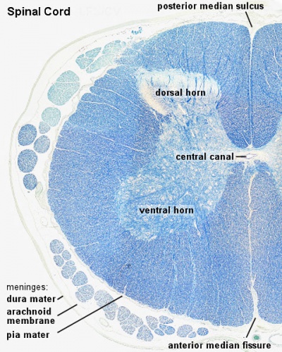
Diagram of a microscope and label
Fluorescence microscope - Wikipedia A fluorescence microscope is an optical microscope that ... and emission characteristics of the fluorophore used to label the ... design shown in the diagram. The Parts of a Microscope (Labeled) Printable Printable ... The Parts of a Microscope (Labeled) Printable. This diagram labels and explains the function of each part of a microscope. Use this printable as a handout or transparency to help prepare students for working with laboratory equipment. Grade: Microscope Poster - Diagram with Labels - Teach Starter A poster containing a diagram with labels showing the key parts of a microscope. In Science it is important that students know how to use a variety of tools when conducting scientific experiments and inquiry. This poster focuses on the microscope and highlights its key parts. Print on tabloid paper to display around your school's science lab ...
Diagram of a microscope and label. PDF Label parts of the Microscope: Answers Label parts of the Microscope: Answers Coarse Focus Fine Focus Eyepiece Arm Rack Stop Stage Clip . Created Date: 20150715115425Z ... Labeling the Parts of the Microscope | Microscope World ... Labeling the Parts of the Microscope This activity has been designed for use in homes and schools. Each microscope layout (both blank and the version with answers) are available as PDF downloads. You can view a more in-depth review of each part of the microscope here. Download the Label the Parts of the Microscope PDF printable version here. Neuron under Microscope with Labeled Diagram ... Neuron under Microscope with Labeled Diagram 31/03/2022 31/03/2022 by anatomylearner The structural and functional unit of the nervous system is the neuron that may easily observe under a light microscope. Neurons may vary considerably in size, shape, and other features. Label Microscope Diagram - EnchantedLearning.com Label Microscope Diagram Using the terms listed below, label the microscope diagram. Inventions and Inventors: arm - this attaches the eyepiece and body tube to the base. base - this supports the microscope. body tube - the tube that supports the eyepiece. coarse focus adjustment - a knob that makes large adjustments to the focus.
Simple Microscope - Diagram (Parts labelled), Principle ... Parts of a Simple Microscope A simple microscope consists of Optical parts Mechanical parts Labeled Diagram of simple microscope parts Optical parts The optical parts of a simple microscope include Lens Mirror Eyepiece Lens A simple microscope uses biconvex lens to magnify the image of a specimen under focus. Compound Microscope- Definition, Labeled Diagram ... The optical microscope often referred to as the light microscope, is a type of microscope that uses visible light and a system of lenses to magnify images of small subjects. There are two basic types of optical microscopes: Simple microscopes. Compound microscopes. The term "compound" in compound microscopes refers to the microscope having ... A Study of the Microscope and its Functions With a Labeled Diagram These labeled microscope diagrams and the functions of its various parts, attempt to simplify the microscope for you. However, as the saying goes, 'practice makes perfect', here is a blank compound microscope diagram and blank electron microscope diagram to label. Download the diagrams and practice labeling the different parts of these ... Welcome to Virtual Urchin - University of Washington Major update Apr 2021: All of the activities on the site are now mobile compatible !! Computers are still recommended, and tablets are preferable to phones: please read the Notes at the bottom of this page for details on the latest updates, mobile compatibility and general information about using this site.
Label Cell Parts | Plant & Animal Cell Activity | StoryboardThat Have your students label a plant and animal cell using one of the landscape poster layouts (small or large). Students will create a cell diagram labeled with the different organelles of plant and animal cells. The cell diagrams are easily colorable, allowing students to differentiate the different parts of the plant and animal cell quickly. Compound Microscope Parts - Labeled Diagram and their ... There are three major structural parts of a compound microscope. The head includes the upper part of the microscope, which houses the most critical optical components, and the eyepiece tube of the microscope. The base acts as the foundation of microscopes and houses the illuminator. The arm connects between the base and the head parts. Connective Tissue and Quiz 1 - histology Note at high power that fibrils or fibers of any type cannot be readily observed. Now with slide 28 (make sure your slide is stained with silver; it should say “Ag” on the label!), note how a network of fine black fibrils is present in this same tissue following silver staining. These are reticular fibers, found in skin, muscle and blood ... Parts of Stereo Microscope (Dissecting microscope ... Labeled part diagram of a stereo microscope Major structural parts of a stereo microscope. There are three major structural parts of a stereo microscope. The viewing Head includes the upper part of the microscope, which houses the most critical optical components, including the eyepiece, objective lens, and light source of the microscope.
ANAT2511 Introduction to Histology - Embryology
Parts Of The Microscope Label Worksheets & Teaching ... Learn the parts of a microscope with this resource!Included in this resource are two pdf documents. One is an answer key/review sheet of a labeled microscope. The other is the microscope with the label boxes blank that I used as the 'quiz'. (Files include a link to editable doc, so you can rewrite a
Quia - Chapter 3: Bacteria & Protists
What is a Bunsen Burner? | Bunsen Burner Parts, Diagram ... Jul 29, 2021 · The Parts of a Bunsen Burner. The entire burner is typically made of metal and it burner sits on the bench via a sturdy base. The main gas inlet at the base of the burner is attached to gas nozzle ...

BodyPartChart Simplified Muscular System - Labeled - Anatomical Charts
Microscope Types (with labeled diagrams) and Functions Simple microscope labeled diagram Simple microscope functions It is used in industrial applications like: Watchmakers to assemble watches Cloth industry to count the number of threads or fibers in a cloth Jewelers to examine the finer parts of jewelry Miniature artists to examine and build their work Also used to inspect finer details on products

Roots, Stems and Leaves Diagrams
Microscope, Microscope Parts, Labeled Diagram, and Functions Microscope, Microscope Parts, Labeled Diagram, and Functions What is Microscope? A microscope is a laboratory instrument used to examine objects that are too small to be seen by the naked eye. It is derived from Ancient Greek words and composed of mikrós, "small" and skopeîn,"to look" or "see".

Leukocytes. Causes, symptoms, treatment Leukocytes
Microscope labeled diagram - SlideShare Microscope labeled diagram 1. The Microscope Image courtesy of: Microscopehelp.com Basic rules to using the microscope 1. You should always carry a microscope with two hands, one on the arm and the other under the base. 2. You should always start on the lowest power objective lens and should always leave the microscope on the low power lens ...
Histology A464
PDF Parts of a Microscope Printables - Homeschool Creations Label the parts of the microscope. You can use the word bank below to fill in the blanks or cut and paste the words at the bottom. Microscope Created by Jolanthe @ HomeschoolCreations.net eyepiece head objective lenses arm focusing knob base illuminator stage stage clips nosepiece.
Angiosperms Biology 2 Notes
Microscope Labeling Practice Diagram | Quizlet Where the microscope slide is placed for viewing. Coarse Focus Knob (Coarse Adjustment) Elevates or lowers the stage a large distance with each turn of the knob. Fine Focus Knob (Fine Adjustment) Elevates or lowers the stage a small distance with each turn or the knob. Base Provides a firm and steady support. Carry with one hand here. Body Tube
Search in gallery
Compound Microscope Parts, Functions, and Labeled Diagram Compound Microscope Parts, Functions, and Labeled Diagram Parts of a Compound Microscope Each part of the compound microscope serves its own unique function, with each being important to the function of the scope as a whole.
Respiratory System | histology
Microscope Drawing And Label at PaintingValley.com ... Tags: microscope, label All rights to paintings and other images found on PaintingValley.com are owned by their respective owners (authors, artists), and the Administration of the website doesn't bear responsibility for their use.
Post a Comment for "43 diagram of a microscope and label"