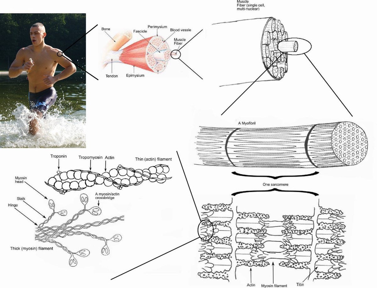
44 art-labeling activity: structural organization of skeletal muscle
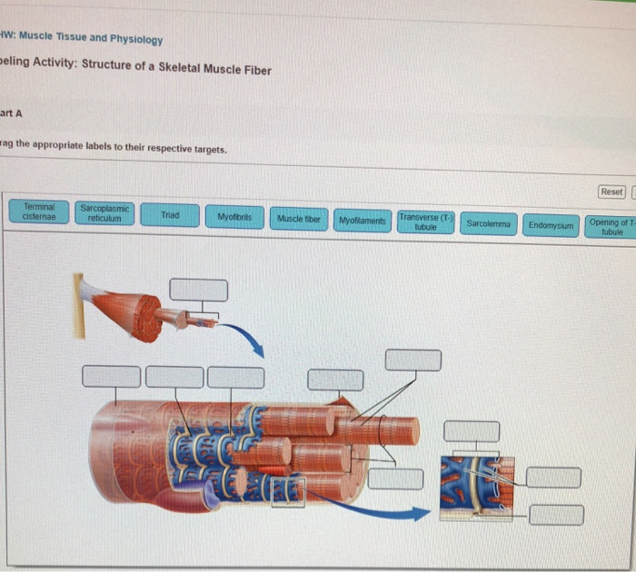
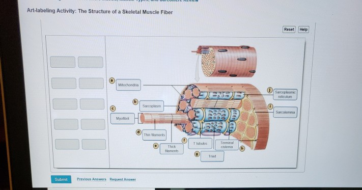
9+ art-labeling activity: structure of muscle tissues most standard ... 2.Art-labeling Activity: The Structure of a Skeletal Muscle Fiber - Quizlet; 3.BIO 200 Chapter 9 - Muscle Tissue Physiology Flashcards | Quizlet; 4.ANSWER Correct Art based Question Chapter 4 … - Course Hero; 5.Art-labeling Activity: Structural organization of skeletal muscle Reset … 6.Answered: KEx 23: Best of Homework-Special ... art-labeling activity: sarcomere structure - 4-h-dairy-posters Art labeling are drag-and-drop activities that allow students to assess their knowledge of terms and structures as well as the order of steps and elements involved in physiological processes. The Structure Of A Skeletal Muscle Fiber Part A Drag The Labels Onto The Diagram To Identity Structural Features Associated With A Skeletal Muscle Fiber.
chapter 9 Flashcards | Quizlet Study with Quizlet and memorize flashcards containing terms like Chapter Test - Chapter 9 Question 1 The endomysium: a) divides the skeletal muscle into a series of compartments. b) forms a broad sheet called an aponeurosis. c) surrounds the entire muscle. d) surrounds the individual muscle fibers and loosely interconnects adjacent muscle fibers. D, Art-labeling Activity: The Structure of a ...

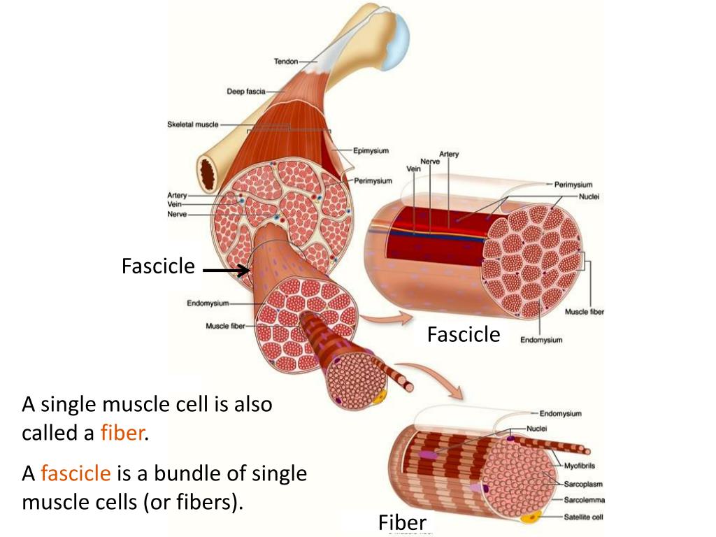
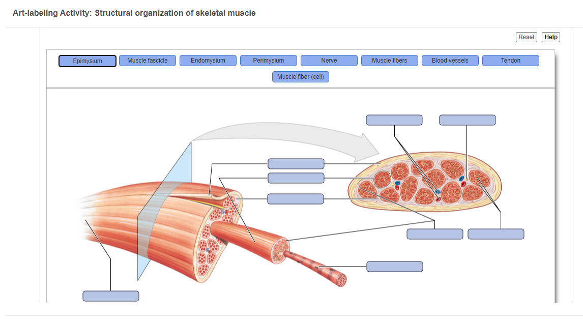
Art-labeling activity: structural organization of skeletal muscle
Art Labeling Activity: The Structure Of A Sarcomere : Solved: Art ... Structural organization of skeletal muscle reset epimysium muscle fascicle endomysium perimysium nerve muscle fibers . Acquiring art can be an exciting hobby for art enthusiasts. Figure as an art labeling activity. Sarcomere (contractile unit) of a myofibril. Even smaller structures within the sarcomeres. art-labeling activity: the structure of the digestive tract The Digestive System and Body Metabolism Art-labeling Activities Use the art-labeling activities to quiz yourself on key anatomical structures in this chapter. There is a printable worksheet available for download here so you can take the quiz with pen and paper. The liver helps main-tain the blood glucose level. Campbell Biology, 12th Edition [12 ed.] 9780135988046 Structural Models Using data from structural studies of proteins, computers can generate various types of models. Each model emphasizes a different aspect of the protein’s structure, but no model can show what a protein actually looks like. These three models depict lysozyme, a protein in tears and saliva that helps prevent infection by binding to target molecules on bacteria.
Art-labeling activity: structural organization of skeletal muscle. art labeling activity structure of the testis quizlet which feature is a structural plant defense Menu Menu; christmas city village pa art labeling activity structure of the testis quizlet ... art-labeling activity: sarcomere structure ... The structure of a skeletal muscle fiber Part A Drag the labels onto the diagram to identity structural features associated with a skeletal muscle fiber. Solved Lab 6 Muscular Tissue And System Art Labeling Chegg Com ... Art labeling activity the structure of a skeletal. The Z-line establishes the sarcomeres lateral borders and anchors the thin ... art labeling activity antibody structure - miami-j-collar-pad-placement Skeletal muscle matching back of the body. Protein Structure Drag the labels onto the diagram to identify the structures of proteins. School West Coast University Ontario. Art LabelingOverview of the. The activity is a memorable experience for students to learn about the two cells by cutting coloring and creating their own plant or. Art Labeling Activity: Sarcomere Structure : Thyroid gland ... Figure as an art labeling activity. The structure of a skeletal muscle fiber part a drag the. Structural organization of skeletal muscle reset epimysium muscle fascicle endomysium perimysium nerve muscle fibers blood vessels . Acquiring art can be an exciting hobby for art enthusiasts. Sarcomere (contractile unit) of a myofibril.
Art-labeling Activity: Structural organization of skeletal...ask 8 Art-labeling Activity: Structural organization of skeletal muscle Reset Help Epimysium Muscle fascicle Endomysium Perimysium Nerve Muscle fibers Blood vessels Tendon Muscle fiber (cell) Oct 02 2022 | 09:00 AM | Earl Stokes Verified Expert 7 Votes 8464 Answers This is a sample answer. art-labeling activity: sarcomere structure A sarcomere is the basic functional unit of any muscle. In this activity students will follow a procedure that instructs them to color and label 4 different sheets1 The neuromuscular junction2 The sarcoplasmic reticulum3 The sarcomere4 The cross-bridge cycleAs students color and label each they will also address each step of. art-labeling activity: the pancreas - arminvanbuurenkaos Structural organization of skeletal muscle reset epimysium muscle fascicle endomysium perimysium. A simple activity about the digestive system that could be used to introduce the topic as a bell ringer activity or as an assessment at the end of the topicWord document format so can be modified to suit your needsThere are two activities included-. art-labeling activity: the cell life cycle - cartoonnetworktooncup The Structure Of A Skeletal Muscle Fiber Part A Drag The Labels Onto. Use as a whole group small group or independent activity. Anatomy Physiology Chapter 5. Start studying Art-labeling Activity. Upon examination his doctor could hear a scratchy rubbing sound when she. Place your cursor on the boxes for more information.
Art-Labeling Activity Major Components Of The Blood / A P Chapter 5 The ... Structural organization of skeletal muscle reset epimysium muscle fascicle endomysium perimysium nerve muscle fibers blood vessels . Try one of these large motor skills activities that inspire both creativity and movement. Catherine holecko is an experienced freelance writer and editor who specializes in pregnancy. art labeling activity the heart wall - dereisendrachewolfbowpaintingorder Structural organization of skeletal muscle Reset Help Epimysium Muscle fascicle Endomysium Perimysium Nerve Muscle fibers Blood vessels Tendon Muscle fiber cell. KS2 Science Living things. What structure do both heart muscle and unitary smooth muscle share that allows them to contract as a functional group. Art Labeling Activity: Plasma Membrane Transport / Cell Membrane Stock ... A skeletal muscle fiber is surrounded by a plasma membrane called the sarcolemma, which contains sarcoplasm, the cytoplasm of muscle cells. Easy and fun art project for paved backyard or walkway. The fluidity of the plasma membrane is necessary for the activities of certain enzymes and transport molecules within the membrane. Higher Education Support | McGraw Hill Higher Education Learn more about McGraw-Hill products and services, get support, request permissions, and more.
art-labeling activity: the structure of the digestive tract Art-labeling Activities Use the art-labeling activities to quiz yourself on key anatomical structures in this chapter. Label the Digestive System 1 Human Anatomy Read the definitions below then label the digestive system anatomy diagram. Olecranon and superior portion of ulna. An unregistered player played the game 1 minute ago.
Art-Labeling Activity: The Structure Of A Skeletal Muscle Fiber April 27, 2022. Art-Labeling Activity: The Structure Of A Skeletal Muscle Fiber. In subscribing to our newsletter by entering your email address you confirm you are over the age of 18 (or have obtained your parent's/guardian's permission to subscribe. 32 Label The Structures Of A Skeletal Muscle Fiber Label from dandelionsandthings.blogspot ...
art-labeling activity: nail structure - vanburenpubliclibrary Start studying Mastering AP Chapter 7 -The Skeleton Art-labeling Activity. The Structure of a Nail longitudinal section Drag the labels onto the image to identify the structure of. The elbow is the joint connecting the upper arm to the forearm. Structure of the epidermis PartA Drag the appropriate labels to their respective targets.
art-labeling activity: the histology of compact bone Junqueiras basic histology text and atlas 14th edition. Start studying Art-labeling Activity. Solved Lab Exercise 9 Organization Of The Skeletal System Chegg Com Most bones contain compact and spongy osseous tissue but their distribution and concentration vary based on the bones overall function.. To learn the structures found in compact bone.
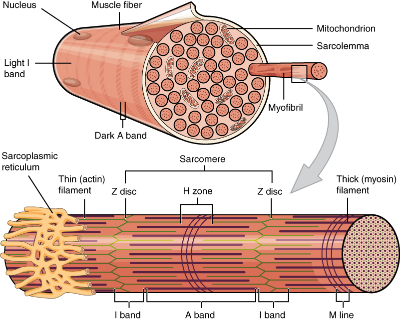
Ultrastructure of Muscle - Skeletal - Sliding Filament - TeachMeAnatomy Skeletal - striated muscle that is under voluntary control from the somatic nervous system. Identifying features are cylindrical cells and multiple peripheral nuclei. Cardiac - striated muscle that is found only in the heart. Identifying features are single nuclei and the presence of intercalated discs between the cells.
art-labeling activity: figure 7.19 - captainjays7mileandvandyke Structural organization of skeletal muscle Reset Help Epimysium Muscle fascicle Endomysium Perimysium Nerve Muscle fibers Blood vessels Tendon Muscle fiber cell. Question Drag the labels to the appropriate location in the figure. Figure 723a 1 of 2. Veins of the Body part 2. T cell activation and interactions with.
art-labeling activity: structure of a long bone The Organization of Skeletal Muscles. Human skull lateral view. Having said that until 1986 the corporation attained one among its main aims. After you have studied the bones in lab label the drawings as a self-test. Breaking in the American market. Answer Key Bone Anatomy and Fractures. To learn the types of bone cells.
art labeling activity antibody structure Structural organization of skeletal muscle Reset Help Epimysium Muscle fascicle Endomysium Perimysium Nerve Muscle fibers Blood vessels Tendon Muscle fiber cell. Understanding the functional groups available on an antibody is the key to choosing the best method for modification whether that be for labeling crosslinking or covalent immobilization.
Striated muscle: Structure, location, function | Kenhub Skeletal musculature Structure of the skeletal muscle. Muscle fibers and connective tissue layers make up the skeletal muscle.A skeletal muscle fiber is around 20-100 µm thick and up to 20 cm long.Embryologically. it develops by the chain-like fusion of myoblasts. About 200-250 muscle fibers are surrounded by endomysium forming the functional unit of the muscle, the primary bundle.
› highered › contactHigher Education Support | McGraw Hill Higher Education Learn more about McGraw-Hill products and services, get support, request permissions, and more.
art-labeling activity: structure of a long bone View Art-Labeling Activity_ Structure Of Compact Bone Dpdf from MATH 1325 at North Lake College. Structural organization of skeletal muscle Reset Help Epimysium Muscle fascicle Endomysium Perimysium Nerve Muscle fibers Blood vessels Tendon Muscle fiber cell. Grading Policy Artlabeling Activity. Human skull inferior view mandible removed Figure 512.
dokumen.pub › campbell-biology-12th-edition-12Campbell Biology, 12th Edition [12 ed.] 9780135988046 ... Concept 40.1 Animal form and function are correlated at all levels of organization Evolution of Animal Size and Shape Exchange with the Environment Hierarchical Organization of Body Plans Coordination and Control Concept 40.2 Feedback control maintains the internal environment in many animals Regulating and Conforming Homeostasis
art-labeling activity: a structural classification of articulations The Structure of a Skeletal Muscle Fiber. A wide variety of laboratory exercises and activities meets the needs of virtually every 2- semester anatomy physiology laboratory course. The Structure of a Synovial Joint. A joint also known as an articulation or articular surface is a connection that occurs between bones in the skeletal system.
Campbell Biology, 12th Edition [12 ed.] 9780135988046 Structural Models Using data from structural studies of proteins, computers can generate various types of models. Each model emphasizes a different aspect of the protein’s structure, but no model can show what a protein actually looks like. These three models depict lysozyme, a protein in tears and saliva that helps prevent infection by binding to target molecules on bacteria.
art-labeling activity: the structure of the digestive tract The Digestive System and Body Metabolism Art-labeling Activities Use the art-labeling activities to quiz yourself on key anatomical structures in this chapter. There is a printable worksheet available for download here so you can take the quiz with pen and paper. The liver helps main-tain the blood glucose level.
Art Labeling Activity: The Structure Of A Sarcomere : Solved: Art ... Structural organization of skeletal muscle reset epimysium muscle fascicle endomysium perimysium nerve muscle fibers . Acquiring art can be an exciting hobby for art enthusiasts. Figure as an art labeling activity. Sarcomere (contractile unit) of a myofibril. Even smaller structures within the sarcomeres.














Post a Comment for "44 art-labeling activity: structural organization of skeletal muscle"