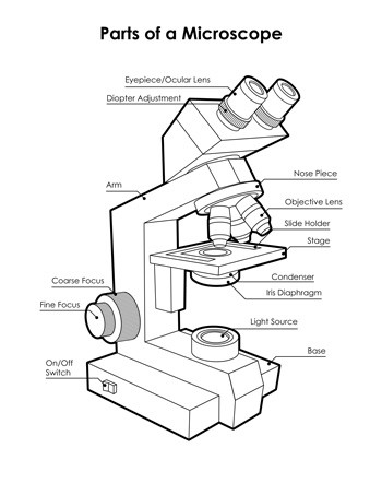
42 labeled diagram of microscope
Labeling the Parts of the Microscope | Microscope World Resources Labeling the Parts of the Microscope This activity has been designed for use in homes and schools. Each microscope layout (both blank and the version with answers) are available as PDF downloads. You can view a more in-depth review of each part of the microscope here. Download the Label the Parts of the Microscope PDF printable version here. Microscope, Microscope Parts, Labeled Diagram, and Functions A wet mount is similar to a sandwich. The slide is the bottom layer. The liquid sample comes next. To minimise evaporation and protect the microscope lens from sample exposure, a small square of clear glass or plastic (a coverslip) is placed on top of the liquid. 1. Collect a clean microscope slide and a coverslip (a thin piece of plastic covering).
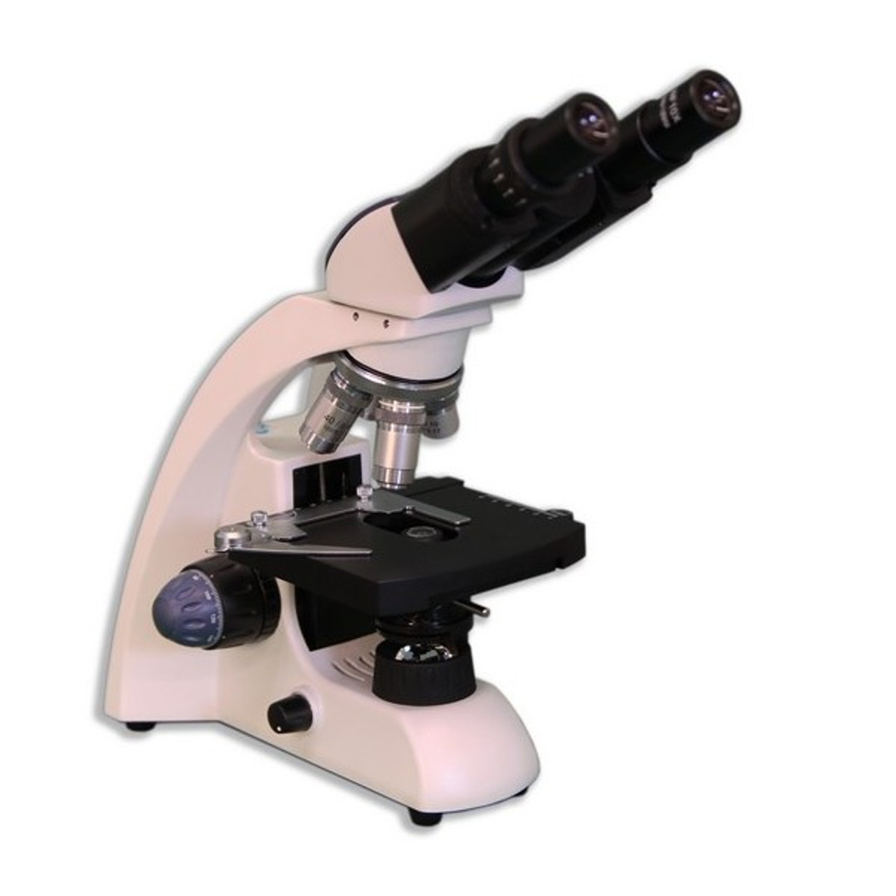
Microscope Labeling - The Biology Corner Students label the parts of the microscope in this photo of a basic laboratory light microscope. Can be used for practice or as a quiz. ... 20. A microscope has an ocular objective of 10x and a high power objective of 50x, what is the microscope's total magnification? _____

Labeled diagram of microscope
anatomylearner.com › dog-skeleton-anatomyDog Skeleton Anatomy with Labeled Diagram - AnatomyLearner Dec 31, 2021 · Here, in the dog skeleton labeled diagram, I tried to show you the different segments of the forelimb, hindlimb with their bones. Again, I tried to show you all the bones from the vertebrae column of a dog skeleton. In addition, in the diagram, you will find a few identified skull bones. The sternum and the ribs are also identified in the dog ... Parts of a Simple Microscope - Labeled (with diagrams) The optical parts of a simple microscope are centered on the specimen - lighting, and magnification. They are as follows: Mirror You can distinguish a simple microscope from other microscopes because of its mirror. It has a plano-convex mirror that focuses the surrounding light on the subject being studied. Lens Light microscopes - Cell structure - Edexcel - BBC Bitesize The magnification of a lens is shown by a multiplication sign followed by the amount the lens magnifies. So a lens magnifying ten times would be ×10. The total magnification of a microscope is:...
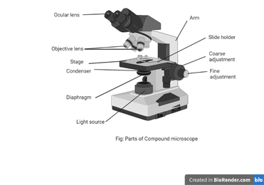
Labeled diagram of microscope. microbenotes.com › parts-of-a-microscopeParts of a microscope with functions and labeled diagram Figure: Diagram of parts of a microscope Head - This is also known as the body. It carries the optical parts in the upper part of the microscope. Base - It acts as microscopes support. It also carries microscopic illuminators. Arms - This is the part connecting the base and to the head and the ... Simple Microscope - Diagram (Parts labelled), Principle, Formula and Uses Labeled Diagram of simple microscope parts Optical parts. The optical parts of a simple microscope include. Lens; Mirror; Eyepiece; Lens. A simple microscope uses biconvex lens to magnify the image of a specimen under focus. Microscope Types (with labeled diagrams) and Functions Compound microscope labeled diagram. Compound microscope functions: It finds great application in areas of pathology, pedology, forensics etc; Its greater order of magnification allows for deeper study of microbial organisms to Detect the cause of diseases; Study the mineral composition in soils; Examine evidences collected in crime scenes by forensics. microscopewiki.com › compound-microscopeCompound Microscope – Diagram (Parts labelled), Principle and ... Feb 03, 2022 · See: Labeled Diagram showing differences between compound and simple microscope parts Structural Components. The three structural components include. 1. Head. This is the upper part of the microscope that houses the optical parts. 2. Arm . This part connects the head with the base and provides stability to the microscope.
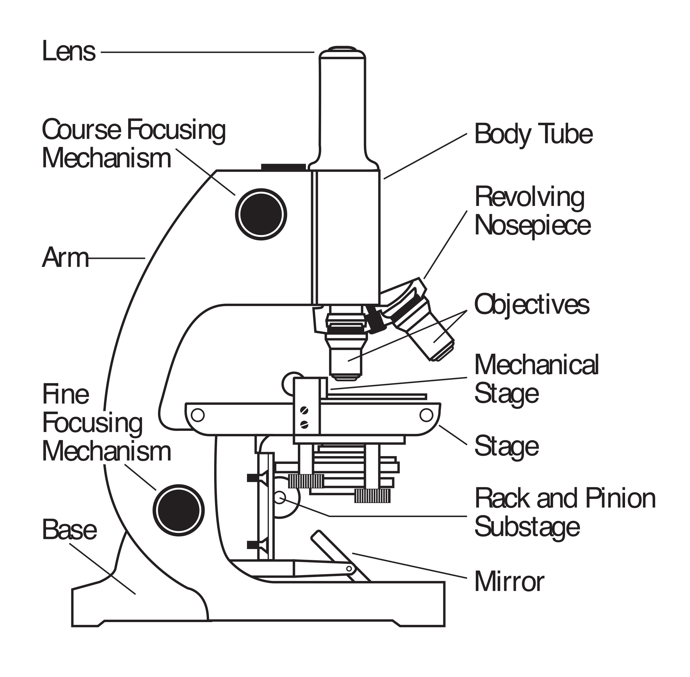
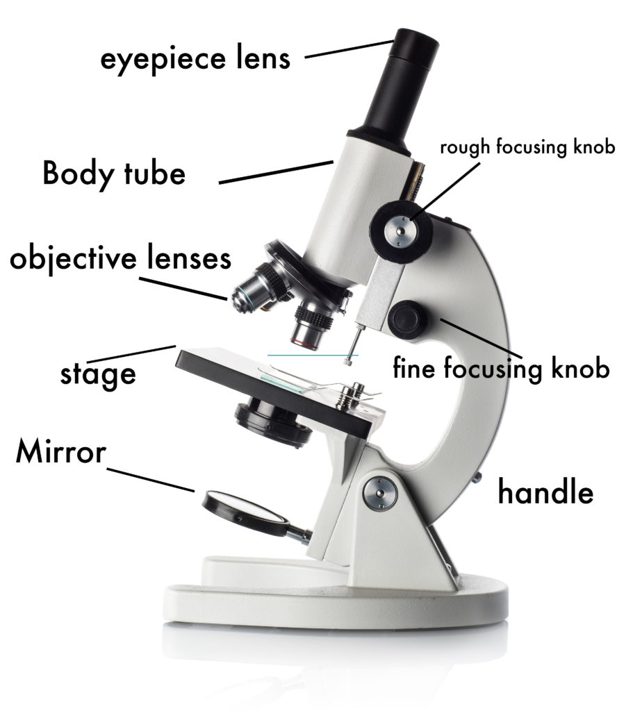
Labelled Diagram of Compound Microscope The below mentioned article provides a labelled diagram of compound microscope. Part # 1. The Stand: The stand is made up of a heavy foot which carries a curved inclinable limb or arm bearing the body tube. The foot is generally horse shoe-shaped structure (Fig. 2) which rests on table top or any other surface on which the microscope in kept. Label Microscope Diagram - EnchantedLearning.com arm - this attaches the eyepiece and body tube to the base. base - this supports the microscope. body tube - the tube that supports the eyepiece. coarse focus adjustment - a knob that makes large adjustments to the focus. diaphragm - an adjustable opening under the stage, allowing different amounts of light onto the stage. microbenotes.com › compound-microscope-principleCompound Microscope- Definition, Labeled Diagram, Principle ... Apr 03, 2022 · Parts of a microscope with functions and labeled diagram Light Microscope- Definition, Principle, Types, Parts, Labeled Diagram, Magnification Amazing 27 Things Under The Microscope With Diagrams anatomylearner.com › cat-skeleton-anatomyCat Skeleton Anatomy with Labeled Diagram - AnatomyLearner May 29, 2021 · Cat skeleton anatomy labeled diagram. Now, I will show you all the bones from the cat skeleton with a diagram. If you find any mistakes in this cat anatomy labeled diagram, please let me know. I hope this cat skeletal system anatomy labeled diagram might help you understand and identify all the cat’s bones.
Microscope Parts and Functions The specimen is placed on the glass and a cover slip is placed over the specimen. This allows the slide to be easily inserted or removed from the microscope. It also allows the specimen to be labeled, transported, and stored without damage. Stage: The flat platform where the slide is placed. microscopewiki.com › electron-microscopeElectron Microscope Principle, Uses, Types and Images ... Feb 02, 2022 · Ans: A light microscope has a low resolving power (0.25µm to 0.3µm) while the electron microscope has a resolution power about 250 times higher than the light microscope at about 0.001µm. Similarly, a light microscope has a magnification of 500X to 1500x while the electron microscope has a much higher magnification of 100,000X to 300,000X. Compound Microscope Parts, Functions, and Labeled Diagram Base: Bottom base of the microscope that houses the illumination & supports the compound microscope. Objective lenses: There are usually 3-5 optical lens objectives on a compound microscope each with different magnification levels. 4x, 10x, 40x, and 100x are the most common magnifying powers used for the objectives. Simple Microscope - Parts, Functions, Diagram and Labelling Parts of the optical parts are as follows: Mirror - A simple microscope has a plano-convex mirror and its primary function is to focus the surrounding light on the object being examined. Lens - The biconvex lens is placed above the stage and its function is to magnify the size of the object being examined.
Free art print of Microscope
Label the Microscope Diagram | Download Scientific Diagram - ResearchGate Download scientific diagram | Label the Microscope Diagram from publication: Laboratory Exercises in Microbiology: Discovering the Unseen World through Hands-on Investigation | Microbiology ...

Microscope Diagram - Label Diagram | Quizlet
Compound Microscope Parts - Labeled Diagram and their Functions Labeled diagram of a compound microscope Major structural parts of a compound microscope There are three major structural parts of a compound microscope. The head includes the upper part of the microscope, which houses the most critical optical components, and the eyepiece tube of the microscope.
Compound Microscope Parts, Functions, and Labeled Diagram ...
Microscope labeled diagram - SlideShare Microscope labeled diagram Oct. 30, 2013 • 6 likes • 27,948 views Download Now Download to read offline Pisgah High School Follow 1. The Microscope Image courtesy of: Microscopehelp.com Basic rules to using the microscope 1. You should always carry a microscope with two hands, one on the arm and the other under the base. 2.
Parts of a Microscope with Their Functions – Microbe Online
Microscope Parts, Function, & Labeled Diagram - slidingmotion Microscope parts labeled diagram gives us all the information about its parts and their position in the microscope. Microscope Parts Labeled Diagram The principle of the Microscope gives you an exact reason to use it. It works on the 3 principles. Magnification Resolving Power Numerical Aperture. Parts of Microscope Head Base Arm Eyepiece Lens

Microscope Parts and Functions
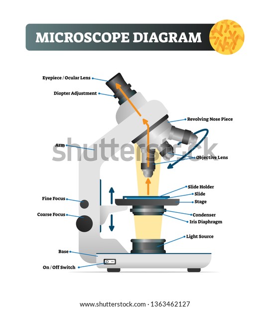
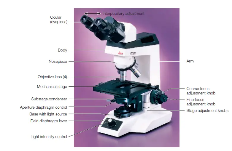
Light Microscope- Definition, Principle, Types, Parts, Labeled Diagram ... Figure: Labeled Diagram of a Light Microscope. Types of light microscopes (optical microscope) With the evolved field of Microbiology, the microscopes. used to view specimens are both simple and compound light microscopes, all using lenses. The difference is simple light microscopes use a single lens for magnification while compound lenses use ...

A labeled diagram of a microscope. MLT 101. :) | Medical lab ...
22 Parts Of a Microscope With Their Function And Labeled Diagram Microscope Description. A microscope is a laboratory instrument used to examine objects that are too small to be seen by the naked eye. In other words, it enlarges images of small objects. Invented by a Dutch spectacle maker in the late 16th century, light microscopes use lenses and light to magnify images.
Microscope Labeled Diagram | Anatomy and Structure
PDF Label parts of the Microscope Label parts of the Microscope: . Created Date: 20150715115425Z

File:Labelledmicroscope.gif - Wikibooks, open books for an ...
Label the microscope Diagram | Quizlet Projects light upwards through the diaphragm, the specimen, and the lenses. Arm. supports the body tube. Stage. Supports the slide being viewed. Coarse Adjustment. focuses the image under low power. Fine Adjustment. sharpens the image under high magnification.
Vektor Stok Microscope Diagram Vector Illustration Labeled ...
Parts of the Microscope with Labeling (also Free Printouts) Parts of the Microscope with Labeling (also Free Printouts) By Editorial Team March 7, 2022 A microscope is one of the invaluable tools in the laboratory setting. It is used to observe things that cannot be seen by the naked eye. Table of Contents 1. Eyepiece 2. Body tube/Head 3. Turret/Nose piece 4. Objective lenses 5. Knobs (fine and coarse) 6.

Label the microscope — Science Learning Hub
Microscope Labeling Diagram | Quizlet Focus and magnify light in differing amounts to view the specimen. Stage Clips. Hold the slide in place on the stage. Nosepiece. Holds the objective lenses and allows the lenses to rotate for viewing. Stage. Supports the slide where the specimen is being viewed. Lamp. Projects or reflects light upward through the diaphragm.

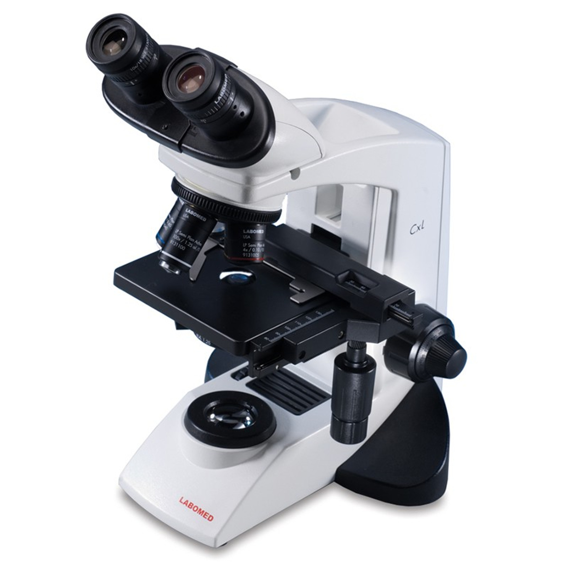
SWIFT M3601 MONOCULAR MICROSCOPE
Microscope Diagram Labeled, Unlabeled and Blank | Parts of a Microscope ... Mar 28, 2016 - Print a microscope diagram, microscope worksheet, or practice microscope quiz in order to learn all the parts of a microscope.
Lab :1
Microscopes - Cell structure - AQA - GCSE Combined Science ... - BBC Late 1600s - Dutch scientist Antonie van Leeuwenhoek constructed a microscope with a single spherical lens. It magnified up to ×275. 1800s - the optical quality of lenses increased and the ...

Compound Microscope- Definition, Labeled Diagram, Principle ...
A Study of the Microscope and its Functions With a Labeled Diagram ... A Study of the Microscope and its Functions With a Labeled Diagram To better understand the structure and function of a microscope, we need to take a look at the labeled microscope diagrams of the compound and electron microscope. These diagrams clearly explain the functioning of the microscopes along with their respective parts.

Diagram of traveling microscope setup with implant cast and ...
Label Microscope Diagram - EnchantedLearning.com Using the terms listed below, label the microscope diagram. arm - this attaches the eyepiece and body tube to the base. base - this supports the microscope. body tube - the tube that supports the eyepiece. coarse focus adjustment - a knob that makes large adjustments to the focus. diaphragm - an adjustable opening under the stage, allowing ...
Microscope - Teaching resources
Label the microscope — Science Learning Hub All microscopes share features in common. In this interactive, you can label the different parts of a microscope. Use this with the Microscope parts activity to help students identify and label the main parts of a microscope and then describe their functions. Drag and drop the text labels onto the microscope diagram. If you want to redo an answer, click on the box and the answer will go back to the top so you can move it to another box.
How to see a plant cell under a compound microscope - Quora
rsscience.com › stereo-microscopeParts of Stereo Microscope (Dissecting microscope) – labeled ... Labeled part diagram of a stereo microscope Major structural parts of a stereo microscope. There are three major structural parts of a stereo microscope. The viewing Head includes the upper part of the microscope, which houses the most critical optical components, including the eyepiece, objective lens, and light source of the microscope.

Compound Microscope Parts, Diagram Definition, Application ...
Light microscopes - Cell structure - Edexcel - BBC Bitesize The magnification of a lens is shown by a multiplication sign followed by the amount the lens magnifies. So a lens magnifying ten times would be ×10. The total magnification of a microscope is:...

Free Microscope Drawing, Download Free Microscope Drawing png ...
Parts of a Simple Microscope - Labeled (with diagrams) The optical parts of a simple microscope are centered on the specimen - lighting, and magnification. They are as follows: Mirror You can distinguish a simple microscope from other microscopes because of its mirror. It has a plano-convex mirror that focuses the surrounding light on the subject being studied. Lens
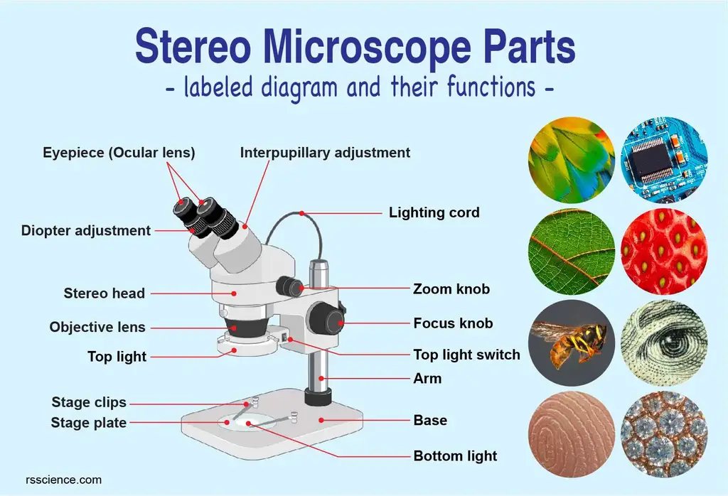
Parts of Stereo Microscope (Dissecting microscope) – labeled ...
anatomylearner.com › dog-skeleton-anatomyDog Skeleton Anatomy with Labeled Diagram - AnatomyLearner Dec 31, 2021 · Here, in the dog skeleton labeled diagram, I tried to show you the different segments of the forelimb, hindlimb with their bones. Again, I tried to show you all the bones from the vertebrae column of a dog skeleton. In addition, in the diagram, you will find a few identified skull bones. The sternum and the ribs are also identified in the dog ...

The Microscope: Create a Labelled Diagram | Teaching Resources

Parts of a Microscope - SmartSchool Systems

Microscope diagram labeled | Clipart Panda - Free Clipart Images

give a well labelled diagram of compound microscope using of ...
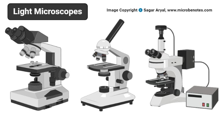
Types of Microscopes and Their Uses – Microbe Online

Light Microscope- Definition, Principle, Types, Parts ...

Compound microscope - their parts and function - Microscopy4kids
Light Microscope- Definition, Principle, Types, Parts ...

Parts of a microscope with functions and labeled diagram

Microscope Maintenance Tips | Science supplies, Multi step ...
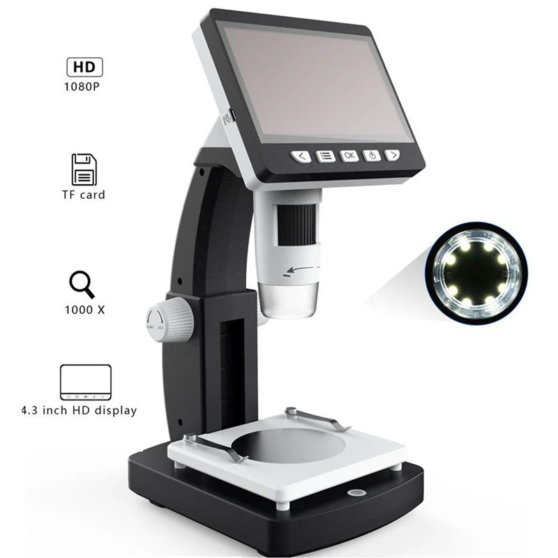
MUSTOOL 1000X Digital Microscope 4.3 inches HD 1080P Portable Desktop LCD Digital Microscope Adjustable 10 Languages 8 LED G710
1.2: Microscopes - Biology LibreTexts

Microscope parts 3D learning APK untuk Unduhan Android

Microscope Labeling Diagram | Quizlet

Lable the microscope worksheet

5 Important Types of Microscopes used in Biology (With Diagram)
Compound Microscope Parts, Functions, and Labeled Diagram ...

Compound and Stereo- microscopes - Microscopes 4 Schools

Compound Microscope Parts – Labeled Diagram and their ...

Microscope, Microscope Parts, Labeled Diagram, and Functions
How to Use a Microscope
Labeled Microscope Diagram - Tim's Printables

microscopy how a microscope works magnification calculations ...
Post a Comment for "42 labeled diagram of microscope"