39 microscope labels and functions
Light Microscope (Theory) - Amrita Vishwa Vidyapeetham Microscope can be separated into optical theory microscopes (Light microscope), electron microscopes (eg.TEM, SEM) and scanning probe microscopes. (eg.AFM, PSTM). Optical microscopes function on the basis of optical theory of lenses by which it can magnifies the image obtained by the movement of a wave through the sample. 18 Instruments used in Microbiology Lab with Principle and Uses List of Instruments used in Microbiology Lab 1. Analytical Balance Working Principle Uses 2. Autoclave Working Principle Uses 3. Bunsen burner Working Principle Uses 4. Centrifuge Working Principle Uses 5. Colony Counter Working Principle Uses 6. Deep Freezer Working Principle Uses 7. Homogenizer Working Principle Uses 8. Hot plate
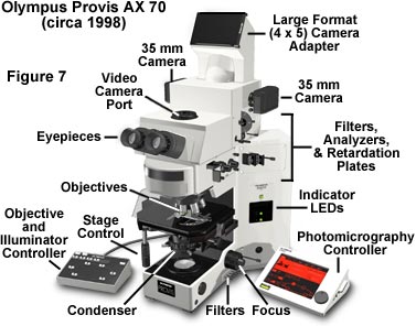
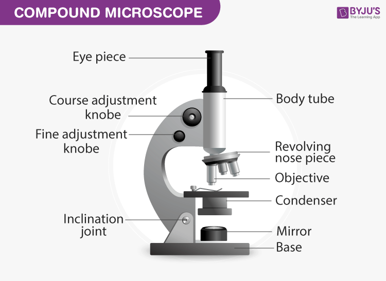
An Introduction to the Light Microscope, Light Microscopy Techniques ... Figure 1: Basic compound microscope: Light from a source is focused onto the sample (object) using a mirror and condenser lens. Light from the sample is collected by an objective, forming an intermediate image which is imaged again by the eyepiece and relayed to the eye, which sees a magnified image of the sample.

Microscope labels and functions
Parts of a Microscope: Lesson for Kids - Study.com Now, look through the microscope like you would a pair of binoculars; these are the ocular lenses ('ocular' means eye). Sometimes when you look it's very dark. Slide your finger down to the base ... Microscope: Types of Microscope, Parts, Uses, Diagram - Embibe A microscope is an optical instrument used to observe and study tiny (microscopic) objects like even cells. The image of an object is magnified through at least one lens in the microscope. This lens bends light toward the eye and makes an object appear larger than it is. Light Microscope- Definition, Principle, Types, Parts, Labeled Diagram ... The functioning of the light microscope is based on its ability to focus a beam of light through a specimen, which is very small and transparent, to produce an image. The image is then passed through one or two lenses for magnification for viewing. The transparency of the specimen allows easy and quick penetration of light.
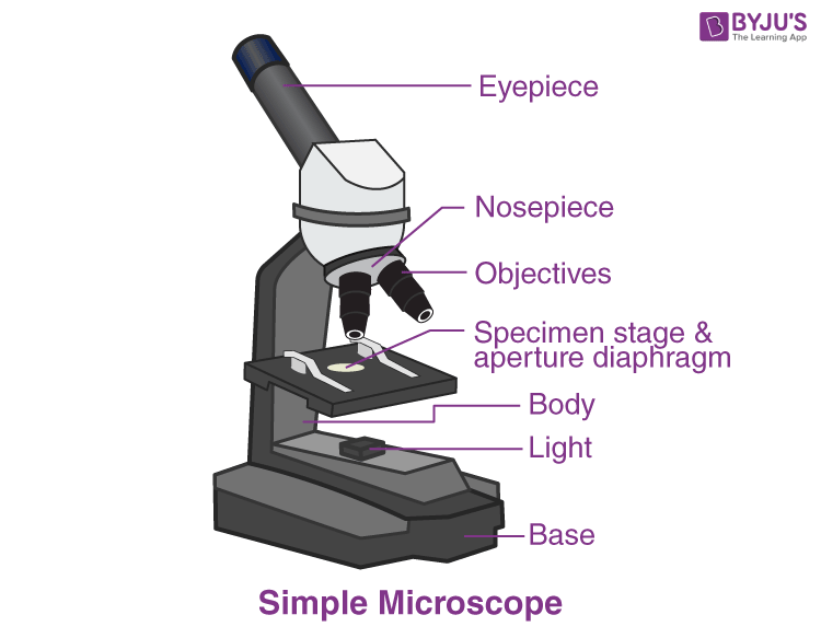
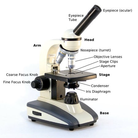
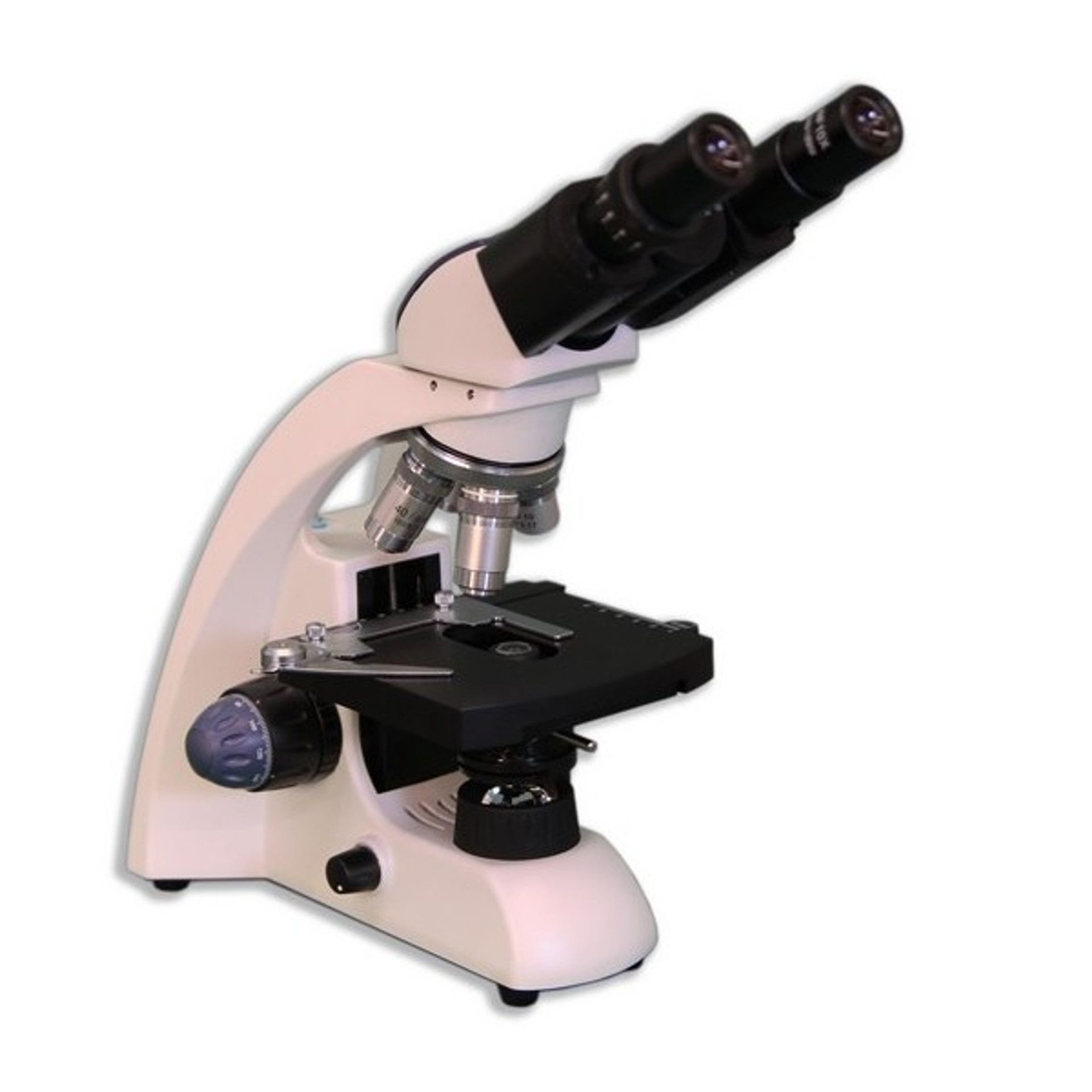
Microscope labels and functions. Simple Microscope - Parts, Functions, Diagram and Labelling Mirror - A simple microscope has a plano-convex mirror and its primary function is to focus the surrounding light on the object being examined. Lens - The biconvex lens is placed above the stage and its function is to magnify the size of the object being examined. Binocular Microscope Anatomy - Parts and Functions with a Labeled ... The microscope's condenser is the structure that collects and focuses light from the light source. You will see an aperture in the condenser that controls the amount of light coming through it. A standard light microscope possesses three to five objective lenses that range in power from 10X to 100X. Compound Microscope - Diagram (Parts labelled), Principle and Uses Compound Microscope - Diagram (Parts labelled), Principle and Uses As the name suggests, a compound microscope uses a combination of lenses coupled with an artificial light source to magnify an object at various zoom levels to study the object. A compound microscope: Is used to view samples that are not visible to the naked eye Microscope: Parts Of A Microscope With Functions And Labeled Diagram. This includes the part of the microscope that allows for observation. It is located at the apex of the microscope. There is an alternate eyepiece having magnifications varying from 5 to 30X. The standard magnification is 10x. Eyepiece tube:The eyepiece tube includes the eyepiece holder. The eyepiece is located just over the objective lens.
Microscope, Microscope Parts, Labeled Diagram, and Functions Microscopes magnify or enlarge small objects such as cells, microbes, bacteria, viruses, microorganisms etc. at a viewable scale for examination and analysis. Microscopes consist of one or more magnification lenses to enlarge the image of the microscopic objects placed in the focal plane. Human Biology Lab Online | Lab 2 - Microscopes, Cell Structure and Function To understand the structure and function of the cell. Step Two. This "Hands On" lab experiment is titled, "The Proper use of the Compound Light Microscope " These virtual exercises simulate how biologist's use microscopes in the lab. Please go to the following links to get all the instructions for doing your experiments. Parts of the Microscope with Labeling (also Free Printouts) Let us take a look at the different parts of microscopes and their respective functions. 1. Eyepiece it is the topmost part of the microscope. Through the eyepiece, you can visualize the object being studied. Its magnification capacity ranges between 10 and 15 times. 2. Body tube/Head It is the structure that connects the eyepiece to the lenses. Scanning Electron Microscope (SEM) Definition. Scanning electron microscope is a classification of electron microscope that uses raster scanning to produce the images of a specimen by scanning using a focused electron beam on the surface of the specimen. An SEM creates magnified images of the specimen by probing along a rectangular area of the specimen with a focused electron beam.
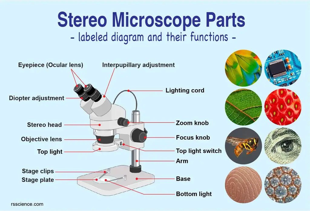
Stereo Microscope - Parts, Types and Uses - Laboratoryinfo.com Stereo Microscope Parts and Functions It has three key parts namely: body, focus block, and viewing head/body. Let us take a look at the functions of every part. Body/viewing head - It houses the optical parts in the upper section of the microscope. Focus block - It attaches the head of the microscope to the stand and focuses it. Light Microscope Parts, Function & Uses - Study.com Anton van Leeuwenhoek (1632-1723) invented a simple (one-lens) microscope around 1670. Leeuwenhoek made lenses by carefully grinding and polishing solid glass to make his microscopes. Parts of a microscope with functions and labeled diagram - Microbe Notes Optical parts of a microscope and their functions The optical parts of the microscope are used to view, magnify, and produce an image from a specimen placed on a slide. These parts include: Eyepiece - also known as the ocular. This is the part used to look through the microscope. Its found at the top of the microscope. Microscopy- History, Classification, Terms, Diagram - The Biology Notes It is simple and can be used to observe living cells and microorganisms. 2 Dark Field Microscopy Dark Field Microscopy uses dark-ground microscopes. The reflected light is used, instead of transmitted light, to form a magnified image. It is used to observe very thin specimens and motility of flagella and cilia. 3 Phase Contrast Microscopy
Electron Microscope Principle, Uses, Types and Images (Labeled Diagram ... Ans: An electron microscope has an evacuated column that is vacuum sealed and houses a cathode, anode, condenser magnet, scatter aperture, specimen chamber, objective lens, fluorescent screen, photographic plate and its transport machinery. This is the reason why the microscope is bulky in size. Q4.
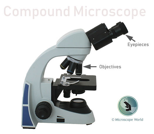
Compound Microscope- Definition, Labeled Diagram, Principle, Parts, Uses The presence or absence of minerals and the presence of metals can be identified using compound microscopes. Students in schools and colleges are benefited from the use of a microscope for conducting their academic experiments. It helps to see and understand the microbial world of bacteria and viruses, which is otherwise invisible to the naked eye.
15 Microscope Parts with Diagram, Location and Function - Study Read A compound microscope has about 15 parts that assist in viewing with a naked eye, a sample holder, a magnifying lens, and a light source. For the convenience of study, we can divide them based on their purpose in the instrument like. A) Parts that assist in viewing the object. B) Part that helps in the adjustment of lenses for a clear view.
Neuron under Microscope with Labeled Diagram - AnatomyLearner Neuron under Microscope with Labeled Diagram. 31/03/2022 31/03/2022 by anatomylearner. The structural and functional unit of the nervous system is the neuron that may easily observe under a light microscope. Neurons may vary considerably in size, shape, and other features. ... According to the function, the neuron may be classified into ...
Microscope Parts, Function, & Labeled Diagram - slidingmotion Microscope parts labeled diagram gives us all the information about its parts and their position in the microscope. Microscope Parts Labeled Diagram The principle of the Microscope gives you an exact reason to use it. It works on the 3 principles. Magnification Resolving Power Numerical Aperture. Parts of Microscope Head Base Arm Eyepiece Lens
Compound Microscope - Types, Parts, Diagram, Functions and Uses Adjustment controls/knob (coarse/fine) - It allows you to easily adjust the focus of the microscope. By adjusting the knob, you can easily increase or decrease the level of details seen when examining at the slide through the eyepiece. Coarse focus - Use this knob with the lowest power objective to get the subject in focus.
Simple Microscope - Diagram (Parts labelled), Principle, Formula and Uses The specimen slide is placed on the stage and is held secure by a pair of clips. Nosepiece It is a revolving turret that houses two or more objective lenses that are used to achieve varying degrees of magnification depending on the need. This nosepiece is a commoncomponent that is seen on all modern day simple and compound microscopes. Base
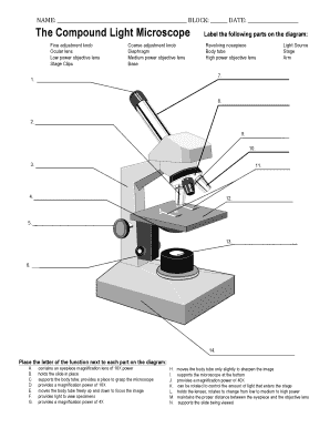
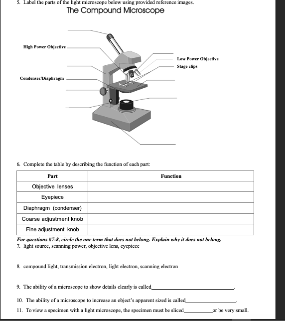
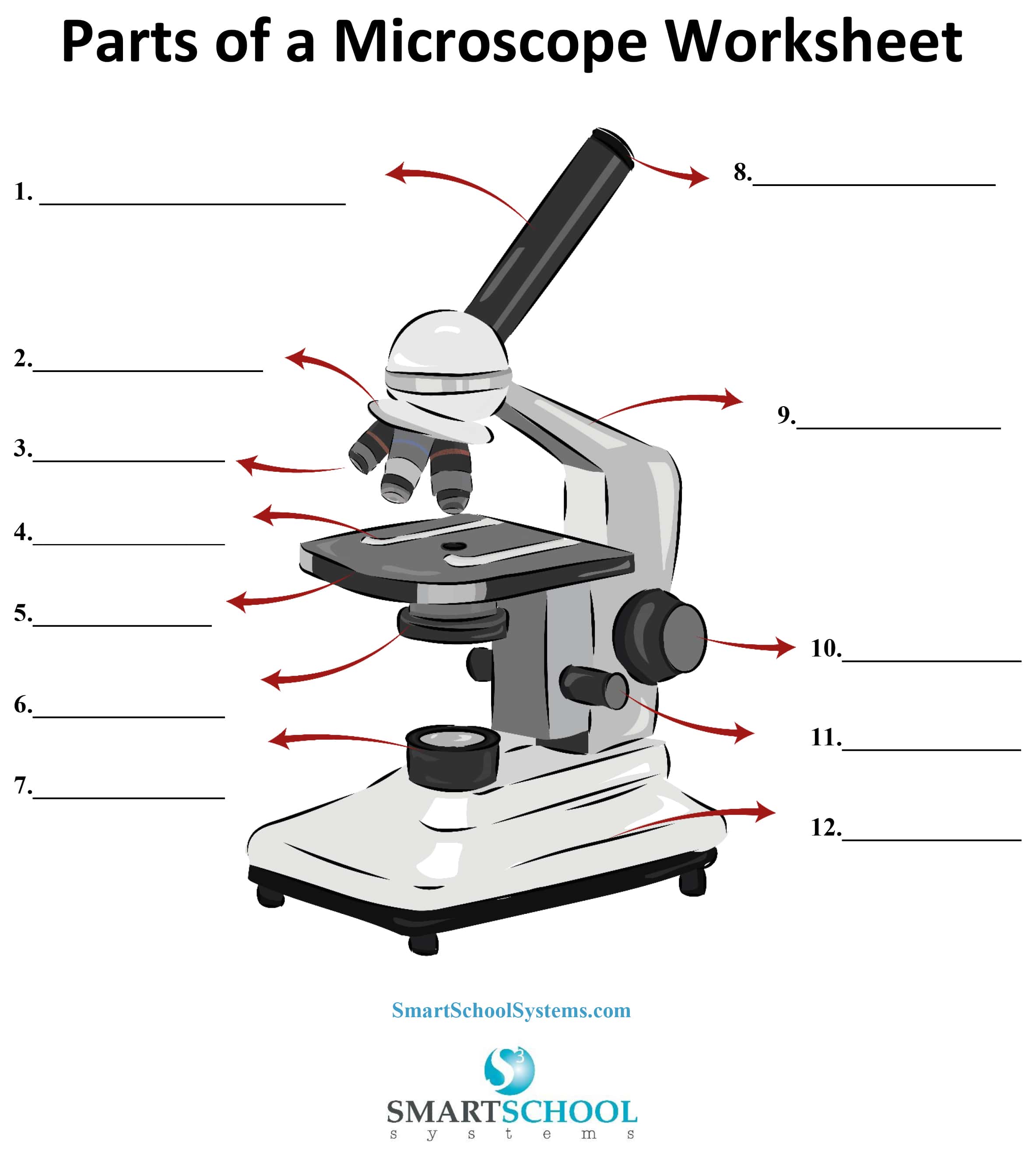
Label The Parts Of A Microscope Worksheet Answers You can use the word bank below to fill in the blanks or cut. Label the parts of a microscope worksheet answers. Students label the parts of the microscope in this photo of a basic laboratory light microscope. Files include a link to editable doc so you can rewrite a. Power 10 x 4 40 Power 10 x 10 100 Power 10 x 40 400 What happens as the power ...
Types of Microscopes - Laboratoryinfo.com Inverted microscope - It is used in biology setting and comes with a light on top and objective lenses beneath the stage. It is specially created for transmitted light observations. Its magnification powers are 40x, 100x, 200x, and 400x. Image 9: The image is one of the classic models of a metallurgical microscope.
Light microscope parts and functions pdf - Australia manuals Working ... Invented by a Dutch spectacle maker in the late 16th century, light microscopes use lenses and light to magnify images. Although a magnifying glass technically qualifies as a simple light microscope, today's high-power—or compound— microscopes use two sets of lenses to give users a much higher level of magnification, … Leica DM750 User Manual 3.
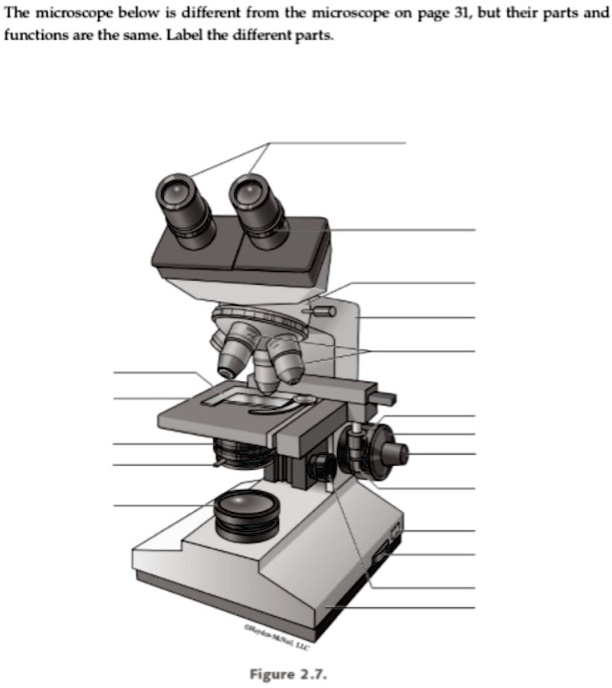
the microscope below i different from the micoscope on page 31 but their parts and functions are the sme label the different parts figure 27 93814
Brightfield Microscope (Compound Light Microscope)- Definition ... The functioning of the microscope is based on its ability to produce a high-resolution image from an adequately provided light source, focused on the image, producing a high-quality image. The specimen which is placed on a microscopic slide is viewed under oil immersion or/and covered with a coverslip. Parts of Brightfield Microscope
Microscope Quiz: How Much You Know About Microscope Parts And Functions ... This quiz will check how much do you know about Microscope Parts and Functions! The microscope has been used in science to understand elements, diseases, and cells. You must have used a microscope back in high school in the biology lab. Do you believe you understood how to use it? Take up the test and see. Questions and Answers 1. Arm: A.
Light Microscope- Definition, Principle, Types, Parts, Labeled Diagram ... The functioning of the light microscope is based on its ability to focus a beam of light through a specimen, which is very small and transparent, to produce an image. The image is then passed through one or two lenses for magnification for viewing. The transparency of the specimen allows easy and quick penetration of light.
Microscope: Types of Microscope, Parts, Uses, Diagram - Embibe A microscope is an optical instrument used to observe and study tiny (microscopic) objects like even cells. The image of an object is magnified through at least one lens in the microscope. This lens bends light toward the eye and makes an object appear larger than it is.
Parts of a Microscope: Lesson for Kids - Study.com Now, look through the microscope like you would a pair of binoculars; these are the ocular lenses ('ocular' means eye). Sometimes when you look it's very dark. Slide your finger down to the base ...

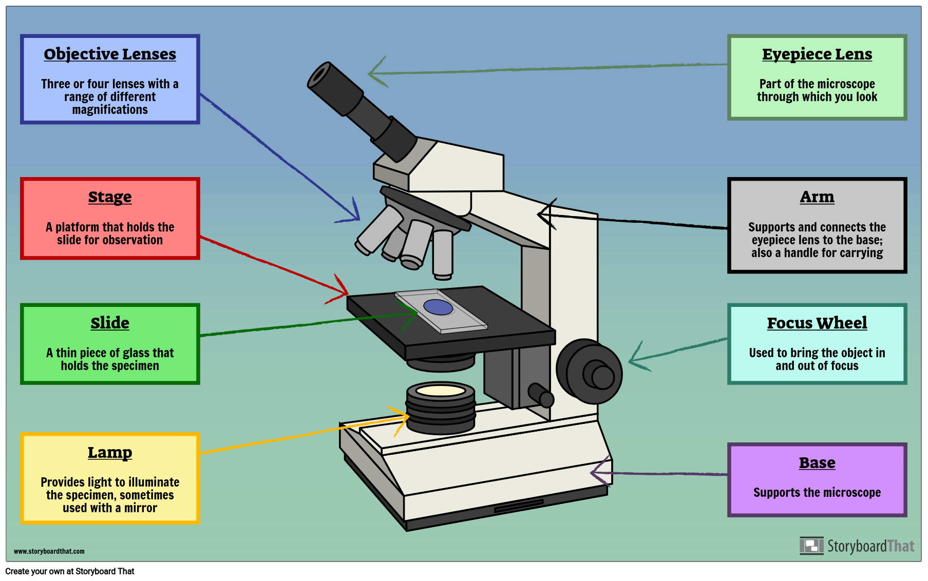
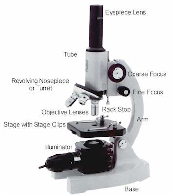

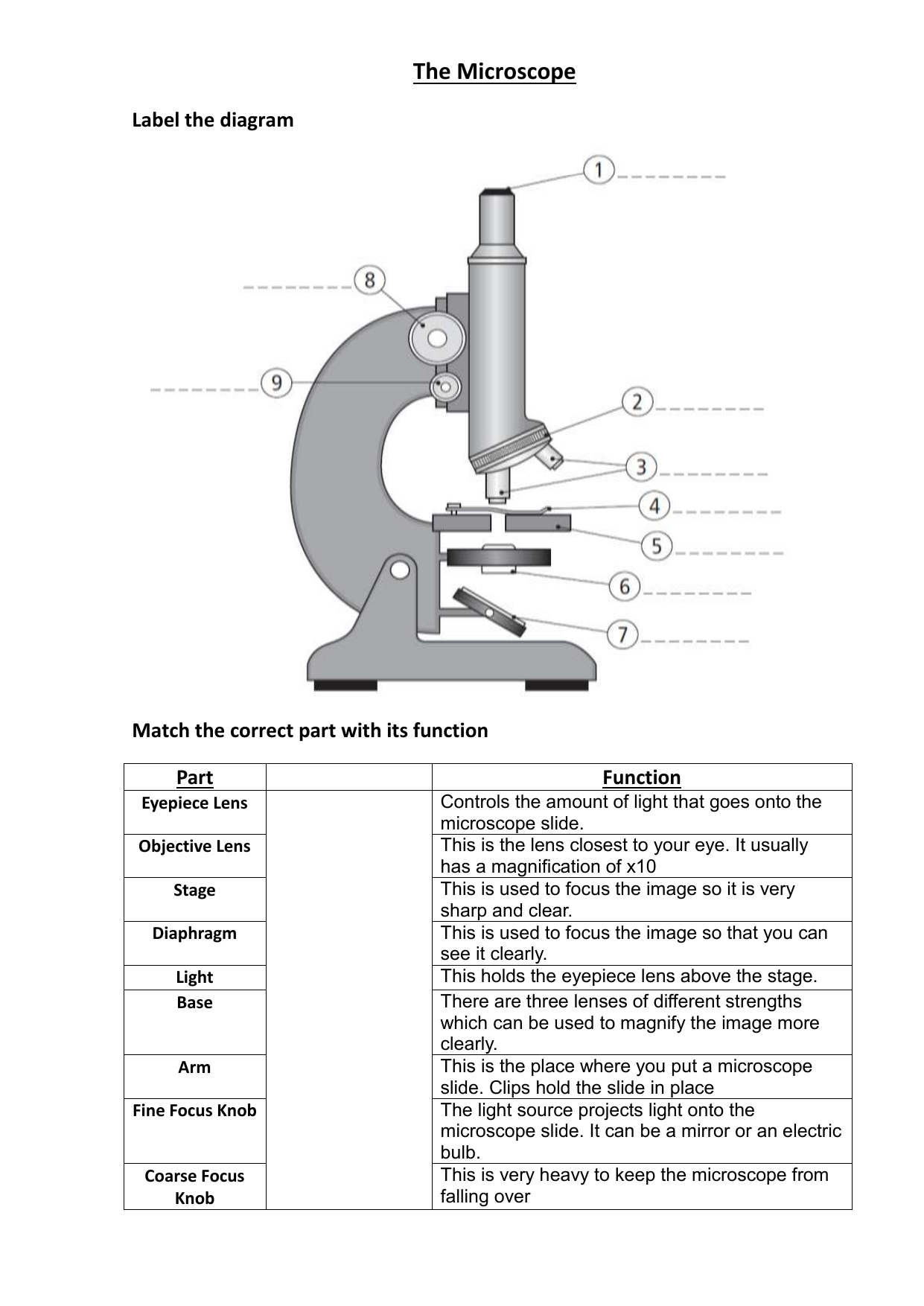













Post a Comment for "39 microscope labels and functions"