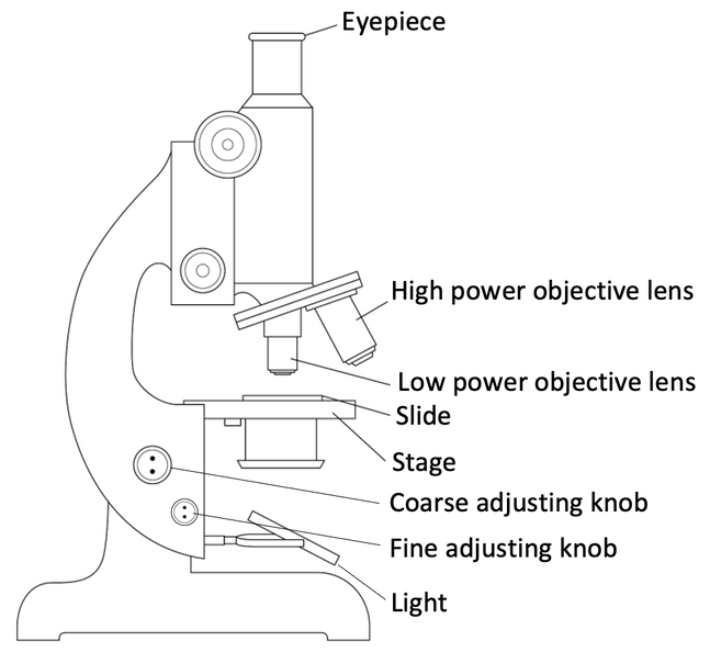
41 light microscope drawing with label
Lesson 1.3 The light microscope - Imago Education Learning Activity 1. Answer the following questions in your exercise book. Study Fig. 1.3 on page 3. This shows the parts of a light microscope. Draw your own light microscope and label the parts. Use the diagram to show how the microscope works (i.e. add notes to the diagram). Compound Microscope: Definition, Diagram, Parts, Uses, Working ... - BYJUS A compound microscope is defined as. A microscope with a high resolution and uses two sets of lenses providing a 2-dimensional image of the sample. The term compound refers to the usage of more than one lens in the microscope. Also, the compound microscope is one of the types of optical microscopes. The other type of optical microscope is a ...
Required practical - using a light microscope - BBC Bitesize Drawing the image Record the microscope images using labelled diagrams or produce digital images. When first examining cells or tissues with low power, draw an image at this stage, even if going ...

Light microscope drawing with label
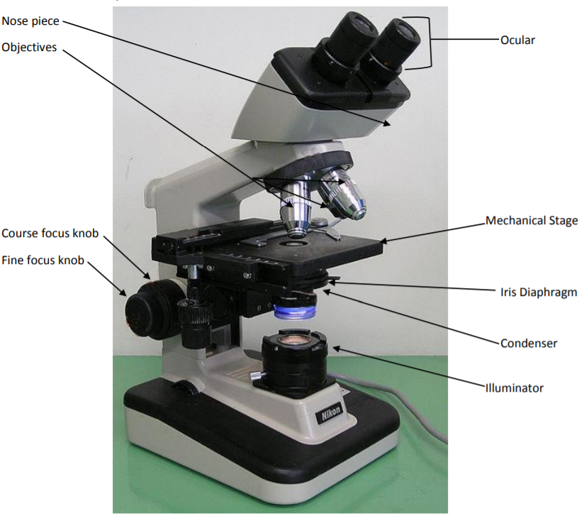
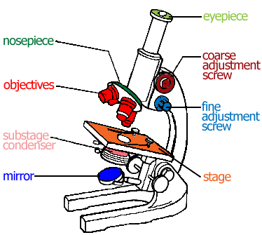
Light microscopes - Cell structure - Edexcel - BBC Bitesize The components of a light microscope and their functions Calculating the magnification of light microscopes. The compound microscope uses two lenses to magnify the specimen: the eyepiece and an ... Compound Microscope - Diagram (Parts labelled), Principle and Uses A compound microscope: Is used to view samples that are not visible to the naked eye. Uses two types of lenses - Objective and ocular lenses. Has a higher level of magnification - Typically up to 2000x. Is used in hospitals and forensic labs by scientists, biologists and researchers to study microorganisms. Microscope Parts, Function, & Labeled Diagram - slidingmotion Condenser. The condenser is to focus the light, which passes from the microscopic illuminator to the specimen. This condenser is located just below the diaphragm. This diaphragm is one of the important parts of the compound microscope which will help to get an accurate and sharp image. The condenser has a magnification power of 400X and above.
Light microscope drawing with label. Microscope Drawing: How to Sketch Microscope Slides Outline the general shapes: Draw the outline of largest shape onto the paper, making it fit within the quarters. Keep you pencil drawings light and adjust the shape as needed. This may require going between the microscope slide and the drawing in order to get the proportions and shape correct. Now move to the other shapes in your field of view. Parts of the Microscope with Labeling (also Free Printouts) 5. Knobs (fine and coarse) By adjusting the knob, you can adjust the focus of the microscope. The majority of the microscope models today have the knobs mounted on the same part of the device. Image 5: The circled parts of the microscope are the fine and coarse adjustment knobs. Picture Source: bp.blogspot.com. › Subjects › animalsAnimal Cell Anatomy & Diagram - Enchanted Learning The cell is the basic unit of life. All organisms are made up of cells (or in some cases, a single cell). Most cells are very small; in fact, most are invisible without using a microscope. Cells are covered by a cell membrane and come in many different shapes. The contents of a cell are called the protoplasm. Glossary of Animal Cell Terms: Cell ... › iet › microscopeVirtual Microscope - NCBioNetwork.org Lesson Description BioNetwork’s Virtual Microscope is the first fully interactive 3D scope - it’s a great practice tool to prepare you for working in a science lab. Explore topics on usage, care, terminology and then interact with a fully functional, virtual microscope.
How to draw compound of Microscope easily - step by step - YouTube I will show you " How to draw compound of microscope easily - step by step "Please watch carefully and try this okay.Thanks for watching.....#microscopedrawi... Compound Microscope Parts, Functions, and Labeled Diagram Common compound microscope parts include: Eyepiece (ocular lens) with or without Pointer: The part that is looked through at the top of the compound microscope. Eyepieces typically have a magnification between 5x & 30x. Monocular or Binocular Head: Structural support that holds & connects the eyepieces to the objective lenses. Light Microscope: Functions, Parts and How to Use It The function of the light microscope is based on its ability to focus a beam of light through a very small and transparent specimen, to produce an image. The image is then passed through one or two lenses for magnification to view. The transparency of the specimen allows for easy and fast light penetration. Specimens can vary from bacteria to ... Pin on Science worksheets - Pinterest Download Clker's Microscope With Labels clip art and related images now. Multiple sizes and related images are all free on Clker.com. A diagram showing all of the parts of a compound light microscope. Mar 7, 2021 - This Pin was discovered by Gloria Legg. Discover (and save!) your own Pins on Pinterest.
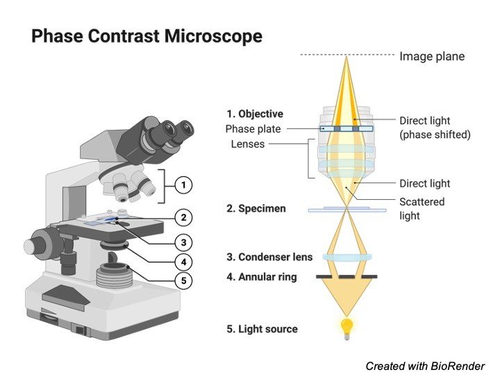
› pmc › articlesSuper resolution fluorescence microscopy - PMC Because microscope objectives collect light from only one side of the sample, a large amount of light is lost, leading to a PSF that is elongated in the axial direction. One can imagine that if the full spherical angle could be covered by the objective, the PSF would become symmetric with the axial size being the same as the lateral size. › products › microscopeMicroscope Imaging Software | Products | Leica Microsystems Jun 15, 2021 · A range of specific modules allows configuration of the microscope as a dedicated high-performance tool for almost any application. The latest software platform, LAS X, encompasses all microscope solutions for Life Science and Industry applications, offering maximum flexibility. The previous Leica Application Suite continues to be supported. Compound Light Microscope Labeling - Printable - PurposeGames.com About this Worksheet. This is a free printable worksheet in PDF format and holds a printable version of the quiz Compound Light Microscope Labeling.By printing out this quiz and taking it with pen and paper creates for a good variation to only playing it online. Animal Cell Under Light Microscope Labelled : Draw and label the ... Animal Cell Under Light Microscope Labelled : Draw and label the diagram of a typical animal plant cell ... - All information about animal cell under microscope labeled.. Add a few drops of a sustainable stain. To prepare a microscope slide of cheek cells, stain them and examine them using a light microscope. Resolving power is the ability to ...
Microscope Drawing posted by Samantha Walker
how to draw microscope (compound) - YouTube drawing microscope. Thank you watching more videos.please subscribe my channel
4: Microscopy - Biology LibreTexts
Microscope, Microscope Parts, Labeled Diagram, and Functions Revolving Nosepiece or Turret: Turret is the part of the microscope that holds two or multiple objective lenses and helps to rotate objective lenses and also helps to easily change power. Objective Lenses: Three are 3 or 4 objective lenses on a microscope. The objective lenses almost always consist of 4x, 10x, 40x and 100x powers. The most common eyepiece lens is 10x and when it coupled with ...

Simple doodles, Microscope parts, Microscopic images
illinois.govIllinois Find places to go, things to see. Search through all the different services offered by the various Illinois agencies.
Microscope, Microscope Parts, Labeled Diagram, and Functions
How to Sketch a Microscope Slide - Identifying and Sketching Cell ... Sketches come to life when you add highlights, shadows and color. For a pencil sketch, separate areas into white, light, medium and dark grey and black. To see the light/dark areas, squint so that the hard edges are blurred and your focus is on the shading. Start shading the light areas by following the shapes.

Microscope Labeling Diagram | Quizlet
learning-center.homesciencetools.com › articleMicroscope Lab Experiments: An Introduction to the Microscope Place one of your homemade slides on the center of the microscope’s stage, directly over the clear hole. Put one stage clip on one edge of the slide to hold it in place leaving the other end free to move around. Turn your microscope’s light source on, lower the stage, and position the lowest power objective lens over the slide.

The Microscope- compound microscope diagram - Major Science ...
Microscope Parts and Functions Microscope Parts and Functions With Labeled Diagram and Functions How does a Compound Microscope Work?. Before exploring microscope parts and functions, you should probably understand that the compound light microscope is more complicated than just a microscope with more than one lens.. First, the purpose of a microscope is to magnify a small object or to magnify the fine details of a larger ...
Microscope Parts and Functions
Labelled Diagram of Compound Microscope - Biology Discussion ADVERTISEMENTS: The below mentioned article provides a labelled diagram of compound microscope. Part # 1. The Stand: The stand is made up of a heavy foot which carries a curved inclinable limb or arm bearing the body tube. The foot is generally horse shoe-shaped structure (Fig. 2) which rests on table top or any other surface on which the ...

Getting to Know the Microscope | Manualzz
Labeling the Parts of the Microscope Labeling the Parts of the Microscope. This activity has been designed for use in homes and schools. Each microscope layout (both blank and the version with answers) are available as PDF downloads. You can view a more in-depth review of each part of the microscope here.
Vektor Stok Vector Microscope (Tanpa Royalti) 1209424708 ...
compound light microscope drawing with label Microscope parts compound ... compound light microscope drawing with label Microscope parts compound diagram drawing light worksheet clipart use sketch answer labeled key label slide 3d simple functions cliparts easy. Xbox games play watchmojo bioshock Polo son Xbox 360; how to get free gamerpics and themes (with download) 6 tips for operating in rainbow six siege like a ...

Label the microscope — Science Learning Hub
Parts of a microscope with functions and labeled diagram Q. Differentiate between a condenser and an Abbe condenser. Ans. Condensers are lenses that are used to collect and focus light from the illuminator into the specimen. They are found under the stage next to the diaphragm of the microscope. They play a major role in ensuring clear sharp images are produced with a high magnification of 400X and above.
Drawing microscope - Teaching resources
Light Microscope- Definition, Principle, Types, Parts, Labeled Diagram ... A light microscope is a biology laboratory instrument or tool, that uses visible light to detect and magnify very small objects and enlarge them. They use lenses to focus light on the specimen, magnifying it thus producing an image. The specimen is normally placed close to the microscopic lens.
Compound Microscope Drawing - ClipArt Best
Label the microscope — Science Learning Hub Use this interactive to identify and label the main parts of a microscope. Drag and drop the text labels onto the microscope diagram. eye piece lens: The lens you look through - normally 10x or 15x magnification. eye piece lens. coarse focus adjustment: Moves the lens up or down and adjusts focus. coarse focus adjustment.

Microscope - Aplikasi di Google Play
Compound Microscope Parts - Labeled Diagram and their Functions - Rs ... There are two major optical lens parts of a microscope: Eyepiece (10x) and Objective lenses (4x, 10x, 40x, 100x). Total magnification power is calculated by multiplying the magnification of the eyepiece and objective lens. The illuminator provides a source of light. The light is focused by the condenser and passing through the specimen placed ...
1 p9 microscope 231110
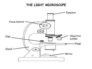
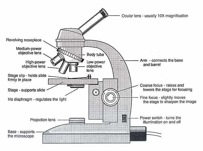
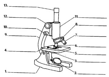
Labeling the Parts of the Microscope | Microscope activity, Science ... Description Worksheet identifying the parts of the compound light microscope. Answer key: 1. Body tube 2. Revolving nosepiece 3. Low power objective 4. Medium power objective 5. High power objective 6. Stage clips 7. Diaphragm 8. Light source 9. Eyepiece 10. Arm 11. Stage 12. Coarse adjustment knob 13. Fine adjustment knob 14. Base
Research Equipment Isolated Biology Microscope Monochrome ...
Labelled Diagram Of A Light Microscope | Products & Suppliers ... Knight Optical (UK) Ltd Custom Plane Mirrors for Microscopes.Plane mirrors also known as front surface mirrors or first surface mirrors are used widely within Microscope applications. As stock we hold a number of general purpose, l/1 and l/4 with a range of up to 6 types of coatings such as Enhanced Aluminium, Ali/SiO2 and Ali/Mgf2.

Parts of a Compound Microscope and Their Functions
vlab.amrita.eduGram Stain Technique - Amrita Vishwa Vidyapeetham Jul 11, 2022 · Drawing a circle on the underside of the slide using a glassware-marking pen may be helpful to clearly designate the area in which you will prepare the smear. You may also label the slide with the initials of the name of the organism on the edge of the slide. Care should be taken that the label should not be in contact with the staining reagents.
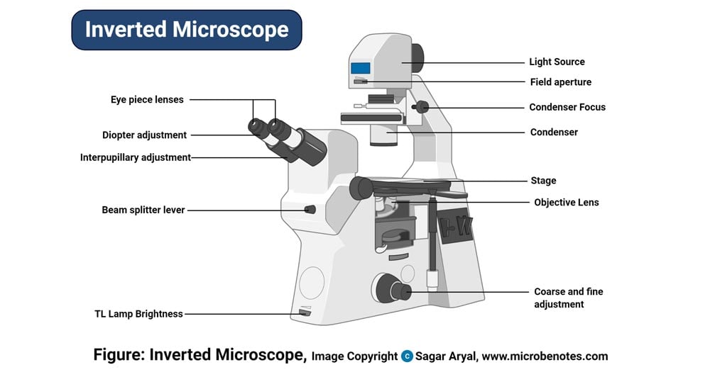
Inverted Microscope- Definition, Principle, Parts, Labeled ...
PDF The Compound Light Microscope The Compound Light Microscope TASK Refer to page 605 in your text to: 1. Name each of the structures described in ... Plug the microscope in, and turn on the power Rotate the nosepiece to the lowest power ... large circle drawn to contain drawing labels are neatly printed labels located on right side of drawing

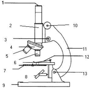
I. Label the parts of a Compound Microscope. You may choose ...
Microscope Parts, Function, & Labeled Diagram - slidingmotion Condenser. The condenser is to focus the light, which passes from the microscopic illuminator to the specimen. This condenser is located just below the diaphragm. This diaphragm is one of the important parts of the compound microscope which will help to get an accurate and sharp image. The condenser has a magnification power of 400X and above.

Compound Microscope Parts – Labeled Diagram and their ...
Compound Microscope - Diagram (Parts labelled), Principle and Uses A compound microscope: Is used to view samples that are not visible to the naked eye. Uses two types of lenses - Objective and ocular lenses. Has a higher level of magnification - Typically up to 2000x. Is used in hospitals and forensic labs by scientists, biologists and researchers to study microorganisms.

Microscope With Labels Clip Art at Clker.com - vector clip ...
Light microscopes - Cell structure - Edexcel - BBC Bitesize The components of a light microscope and their functions Calculating the magnification of light microscopes. The compound microscope uses two lenses to magnify the specimen: the eyepiece and an ...

Living Environment Course

Microscope Diagram Labeled, Unlabeled and Blank | Parts of a ...

Diagram of Compound Microscope

Alternate grade 11 - ccbbiology11
Free Microscope Drawing, Download Free Microscope Drawing png ...
Microscope Drawing - How To Draw A Microscope Step By Step

Old microscope color sketch engraving vector illustration ...
innovative solutions of advanced instrumentation for research ...

How to Draw a Cartoon Microscope – Draw Swan
Cell Structure - Biology | Quiz

Parts of a Microscope Labeling Activity
B1 E) Microscopy: Using Microscopes – AQA Combined Science ...

Simple Microscope - Diagram (Parts labelled), Principle ...

microscopy how a microscope works magnification calculations ...

Microscope Drawing Set stock vector. Illustration of sketch ...

Optical microscope Drawing Worksheet, public space, angle ...

Microscope Diagram Diagram | Quizlet

Microscope diagram labeled | Clipart Panda - Free Clipart Images

Light Microscope PNG - light-microscope-e compound-light ...
Transparent Microscope Vector Png - Cartoon, Png Download ...
Free Microscope Drawing, Download Free Microscope Drawing png ...
Post a Comment for "41 light microscope drawing with label"