39 light microscope diagram with labels
pages.zeiss.com › rs › 896-XMS-794Principles of Fluorescence and Fluorescence Microscopy Figure 6 Basic setup of an inverted widefield fluorescence microscope imaging a sample containing GFP. The first important component is the light source. To produce the optimal wavelength of excitatory light for a particular fluorophore, an excitation filter is inserted in the light path between the light source and the sample. This is the first of Parts of the Microscope with Labeling (also Free Printouts) Parts of the Microscope with Labeling (also Free Printouts) A microscope is one of the invaluable tools in the laboratory setting. It is used to observe things that cannot be seen by the naked eye. Table of Contents 1. Eyepiece 2. Body tube/Head 3. Turret/Nose piece 4. Objective lenses 5. Knobs (fine and coarse) 6. Stage and stage clips 7. Aperture
Pin on Science worksheets - Pinterest A Study of the Microscope and its Functions With a Labeled Diagram To better understand the structure and function of a microscope, we need to take a look at the labeled microscope diagrams of the compound and electron microscope. These diagrams clearly explain the functioning of the microscopes along with their respective parts. M mooketsi

Light microscope diagram with labels
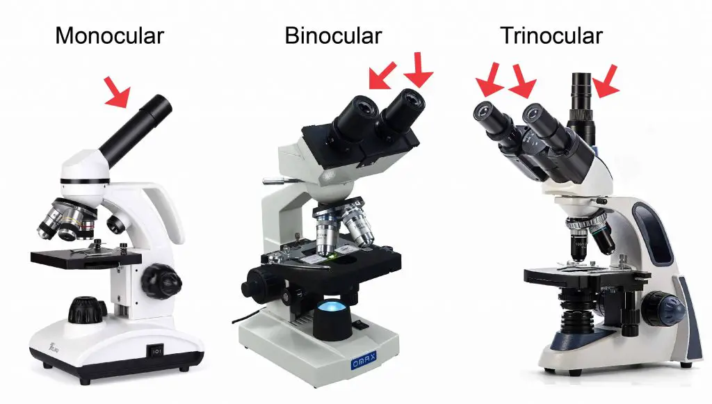
Labelled Diagram Of A Light Microscope | Products & Suppliers ... Products/Services for Labelled Diagram Of A Light Microscope Microscopes - (706 companies) ...and transmission electron microscopes. Acoustic and ultrasonic microscopes use sound waves to create images of the sample. Compound microscopes use a single light path. These types of microscopes can have a single eyepiece (monocular) or a dual eyepiece... › 6-label-the-microscopeLabel the microscope - Science Learning Hub Jun 08, 2018 · All microscopes share features in common. In this interactive, you can label the different parts of a microscope. Use this with the Microscope parts activity to help students identify and label the main parts of a microscope and then describe their functions. Drag and drop the text labels onto the microscope diagram. If you want to redo an ... › en › microscopeFluorescence Resonance Energy Transfer (FRET) Microscopy Presented in Figure 3 is a Jablonski diagram illustrating the coupled transitions involved between the donor emission and acceptor absorbance in fluorescence resonance energy transfer. Absorption and emission transitions are represented by straight vertical arrows (green and red, respectively), while vibrational relaxation is indicated by wavy ...
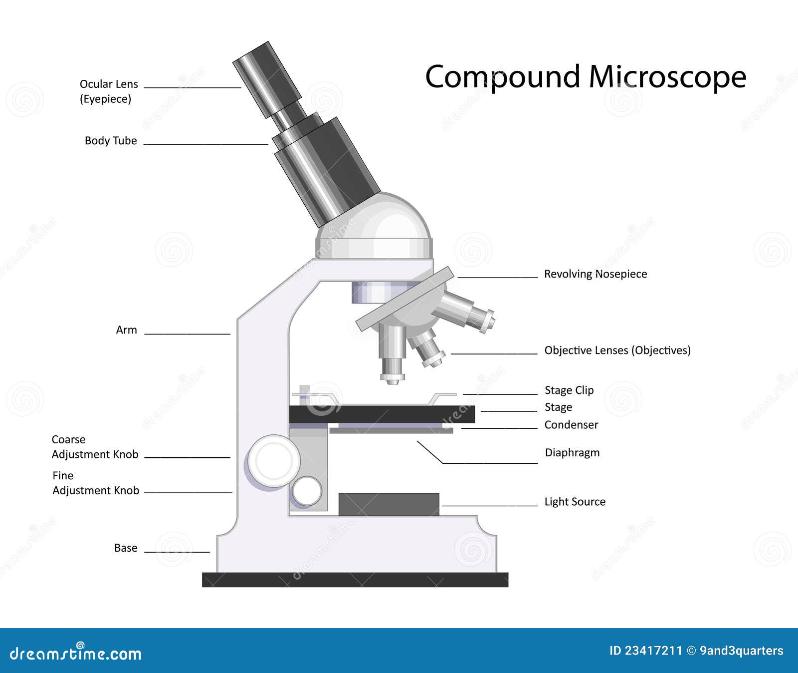
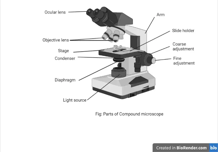
Light microscope diagram with labels. Label a Microscope - Storyboard That Knowing the names of the different parts of the microscope is essential to be able to use one properly. Create a poster that labels the parts of a microscope and includes descriptions of what each part does. Click "Start Assignment". Use a landscape poster layout (large or small). Search for a diagram of a microscope. rsscience.com › stereo-microscopeParts of Stereo Microscope (Dissecting microscope) - Rs' Science Labeled part diagram of a stereo microscope Major structural parts of a stereo microscope. There are three major structural parts of a stereo microscope. The viewing Head includes the upper part of the microscope, which houses the most critical optical components, including the eyepiece, objective lens, and light source of the microscope. Labelled Diagram of Compound Microscope - Biology Discussion The below mentioned article provides a labelled diagram of compound microscope. Part # 1. The Stand: The stand is made up of a heavy foot which carries a curved inclinable limb or arm bearing the body tube. The foot is generally horse shoe-shaped structure (Fig. 2) which rests on table top or any other surface on which the microscope in kept. Microscope Parts and Functions With Labeled Diagram and Functions How ... First, the purpose of a microscope is to magnify a small object or to magnify the fine details of a larger object in order to examine minute specimens that cannot be seen by the naked eye. Here are the important compound microscope parts... Eyepiece: The lens the viewer looks through to see the specimen.
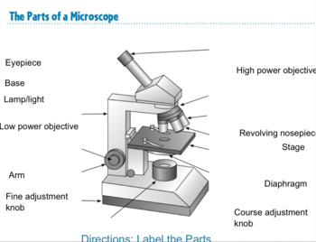
Microscope Labeling - The Biology Corner 1) Start with scanning (the shortest objective) and only use the COARSE knob . Once it is focused… 2) Switch to low power (medium) and only use the COARSE knob . You may need to recenter your slide. Once it is focused.. 3) Switch to high power (long objective). Label the microscope Diagram | Quizlet Start studying Label the microscope. Learn vocabulary, terms, and more with flashcards, games, and other study tools. Compound Microscope Parts, Functions, and Labeled Diagram Compound Microscope Definitions for Labels. Eyepiece (ocular lens) with or without Pointer: The part that is looked through at the top of the compound microscope. Eyepieces typically have a magnification between 5x & 30x. Monocular or Binocular Head: Structural support that holds & connects the eyepieces to the objective lenses. › topics › medicine-andConfocal Microscopy - an overview | ScienceDirect Topics A standards document, which describes confocal microscopy and its influence quantities, has recently completed an ISO ballot as a final draft international standard (ISO FDIS 25178-607, 2018). A schematic diagram of a typical confocal microscope is shown in Fig. 15.1 (ASME B46-2009, 2010; Weller et al., 2012). Most examples of this method rely ...
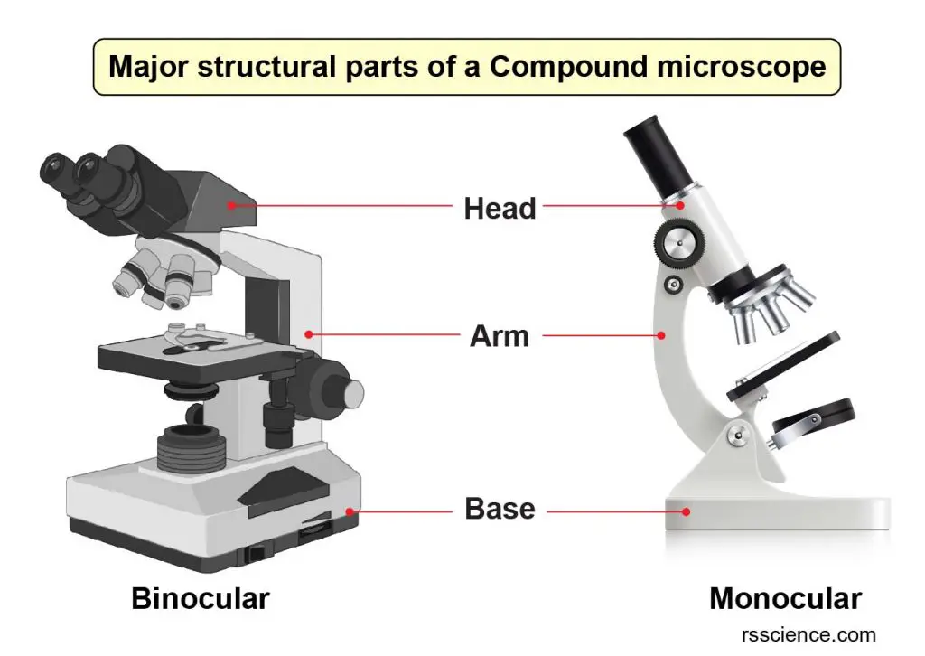
Parts of a microscope with functions and labeled diagram Figure: Diagram of parts of a microscope There are three structural parts of the microscope i.e. head, base, and arm. Head - This is also known as the body. It carries the optical parts in the upper part of the microscope. Base - It acts as microscopes support. It also carries microscopic illuminators. Labeled Microscope and Basics of Life Diagram | Quizlet PLAY. A microscope is an instrument widely to magnify and resolve the image of an object that is otherwise invisible to naked eye. For resolving the details of objects, which otherwise cannot be achieved by naked eye, a microscope is used. This set of flash cards will help the student to identify the different parts and function of the microscope. 22 Parts Of a Microscope With Their Function And Labeled Diagram The field diaphragm control is located around the lens located in the base. Hinge Screw -This screw fixes the arm to the base and allow for the tilting of the arm. Stage Clips - They hold the slide firmly onto the stage. On/OFF Switch - This switch on the base of the microscope turns the illuminator off and on. PDF Label parts of the Microscope Label parts of the Microscope: . Created Date: 20150715115425Z
Parts of a Microscope (Pre-Assessment) - Quizizz
Labeling the Parts of the Microscope | Microscope World Resources Labeling the Parts of the Microscope This activity has been designed for use in homes and schools. Each microscope layout (both blank and the version with answers) are available as PDF downloads. You can view a more in-depth review of each part of the microscope here. Download the Label the Parts of the Microscope PDF printable version here.

Simple Microscope - Diagram (Parts labelled), Principle ...
Label Microscope Diagram - EnchantedLearning.com low-power objective - a small lens with low magnifying power. mirror (or light source) - this directs light upwards onto the slide. revolving nosepiece - the rotating device that holds the objectives (lenses). stage - the platform on which a slide is placed. stage clips - metal clips that hold a slide securely onto the stage.

Microscope - diagram Tom Butler | Science skills, Science ...
Compound Microscope Parts - Labeled Diagram and their Functions - Rs ... The eyepiece (or ocular lens) is the lens part at the top of a microscope that the viewer looks through. The standard eyepiece has a magnification of 10x. You may exchange with an optional eyepiece ranging from 5x - 30x. [In this figure] The structure inside an eyepiece. The current design of the eyepiece is no longer a single convex lens.

Optical Microscope PNG - optical-microscope-parts optical ...
Microscope Labeling - The Biology Corner Students label the parts of the microscope in this photo of a basic laboratory light microscope. Can be used for practice or as a quiz. Name_____ Microscope Labeling . Microscope Use: 15. When focusing a specimen, you should always start with the _____ objective.
Compound Microscope Parts – Labeled Diagram and their ...
Labeling the Parts of the Microscope | Microscope activity, Science ... Description Worksheet identifying the parts of the compound light microscope. Answer key: 1. Body tube 2. Revolving nosepiece 3. Low power objective 4. Medium power objective 5. High power objective 6. ... Print a microscope diagram, microscope worksheet, or practice microscope quiz in order to learn all the parts of a microscope. CCabreza ...
Compound Microscope stock vector. Illustration of research ...
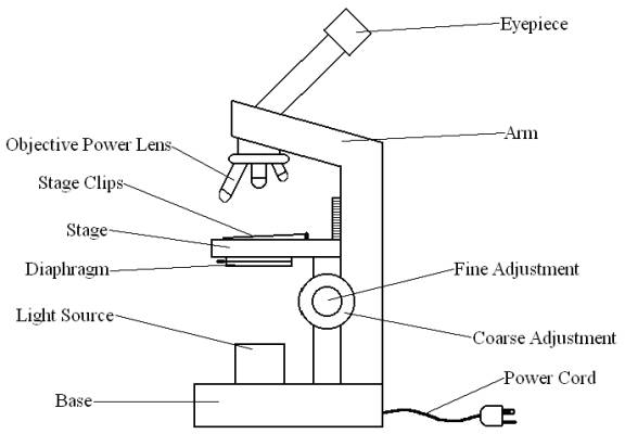
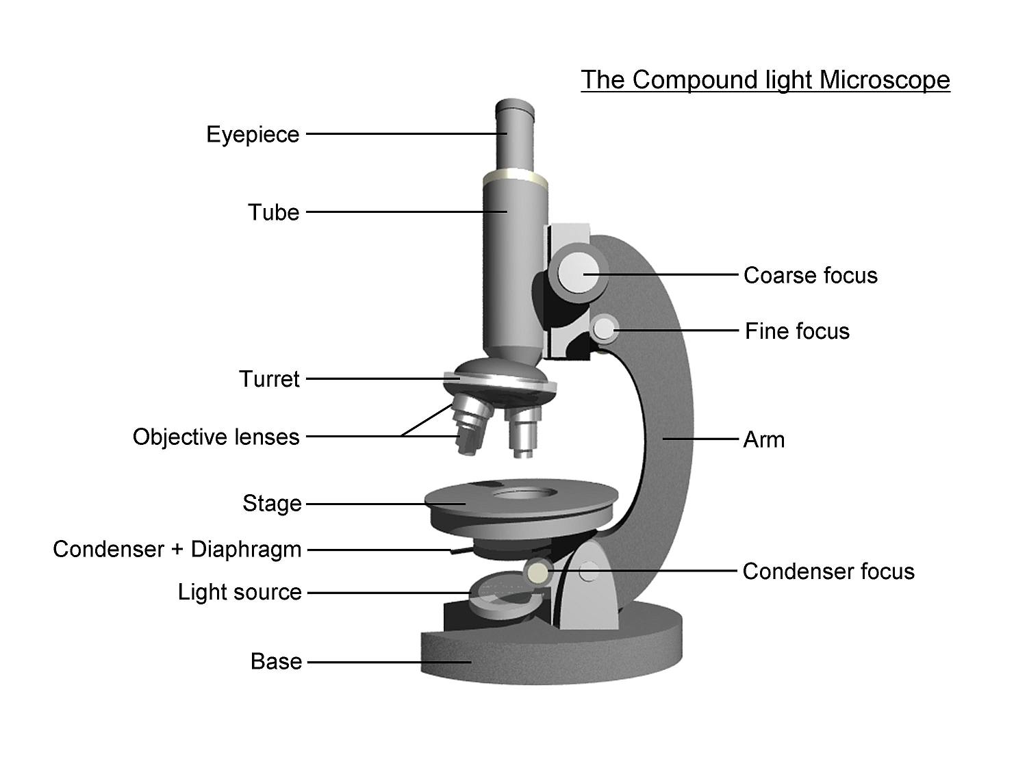
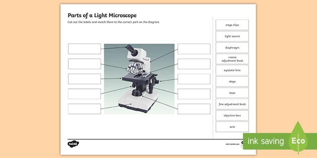
PDF Parts of the Light Microscope - Science Spot Supports the MICROSCOPE D. STAGE CLIPS HOLD the slide in place C. OBJECTIVE LENSES Magnification ranges from 10 X to 40 X F. LIGHT SOURCE Projects light UPWARDS through the diaphragm, the SPECIMEN, and the LENSES H. DIAPHRAGM Regulates the amount of LIGHT on the specimen E. STAGE Supports the SLIDE being viewed K. ARM Used to SUPPORT the
simple light microscope diagram - Clip Art Library
Light Microscope- Definition, Principle, Types, Parts, Labeled Diagram ... A light microscope is a biology laboratory instrument or tool, that uses visible light to detect and magnify very small objects and enlarge them. They use lenses to focus light on the specimen, magnifying it thus producing an image. The specimen is normally placed close to the microscopic lens.

Cytology. Cytology. radiation used to illuminate the specimen ...
Simple Microscope - Diagram (Parts labelled), Principle, Formula and Uses Parts of a Simple Microscope A simple microscope consists of Optical parts Mechanical parts Labeled Diagram of simple microscope parts Optical parts The optical parts of a simple microscope include Lens Mirror Eyepiece Lens A simple microscope uses biconvex lens to magnify the image of a specimen under focus.

label microscope diagram | Charts | Microscope, Anatomy bones ...
Microscope Types (with labeled diagrams) and Functions Simple microscope labeled diagram Simple microscope functions It is used in industrial applications like: Watchmakers to assemble watches Cloth industry to count the number of threads or fibers in a cloth Jewelers to examine the finer parts of jewelry Miniature artists to examine and build their work Also used to inspect finer details on products

Compound Light Microscope Labeling Diagram | Quizlet
PDF COMPOUND LIGHT MICROSCOPE LAB - Springfield Public Schools COMPOUND(LIGHT(MICROSCOPE(LAB((Follow(written(andoral(instructions.((((A.((Label(the(parts(of(the(compound(microscope.(Be(able(to(label(a(blank(diagram

Label the Microscope Diagram | Download Scientific Diagram
en.wikipedia.org › wiki › Two-photon_excitationTwo-photon excitation microscopy - Wikipedia In scattering tissue, on the other hand, the superior optical sectioning and light detection capabilities of the two-photon microscope result in better performance. Applications Main. Two-photon microscopy has been involved with numerous fields including: physiology, neurobiology, embryology and tissue engineering.
Microscope Labeling Activity - SMART Board Activity - Interactive Review
Microscope Parts, Function, & Labeled Diagram - slidingmotion Condenser. The condenser is to focus the light, which passes from the microscopic illuminator to the specimen. This condenser is located just below the diaphragm. This diaphragm is one of the important parts of the compound microscope which will help to get an accurate and sharp image. The condenser has a magnification power of 400X and above.

Labeling the Parts of the Microscope | Microscope World Resources
Microscope, Microscope Parts, Labeled Diagram, and Functions The Iris Diaphragm is located above the condenser lens and below the microscope stage. The different sized holes in the diaphragm helps to vary the size of the cone and intensity of light that is projected upward into the slide. However, there is no set rule regarding which setting to use for a particular power.

Microscope With Labels Clip Art at Clker.com - vector clip ...
PDF Label the Light Microscope Label the Light Microscope: Ocular lens Objective ourse Adjustment Fine Adjustment Light ase Diaphragm rightness control Nosepiece ondenser Mechanical Stage Head with prism Ocular tube Arm. Head Nosepiece Objective turret Stage Iris Diaphragm Illumination System Base Eyepiece ...

Microscope Diagram Labeled, Unlabeled and Blank | Parts of a ...
› confocal-microscopes › lsm-980LSM 980 with Airyscan 2 – Confocal Microscope with Multiplex ... This requires excellent imaging performance combined with low phototoxicity and high speed. LSM 980, your platform for confocal 4D imaging, is optimized for simultaneous spectral detection of multiple weak labels with the highest light efficiency. Employ a wealth of fluorescent labels from 380 nm to 900 nm.

22 Parts Of a Microscope With Their Function And Labeled ...
› en › microscopeFluorescence Resonance Energy Transfer (FRET) Microscopy Presented in Figure 3 is a Jablonski diagram illustrating the coupled transitions involved between the donor emission and acceptor absorbance in fluorescence resonance energy transfer. Absorption and emission transitions are represented by straight vertical arrows (green and red, respectively), while vibrational relaxation is indicated by wavy ...
simple light microscope labeled - Clip Art Library
› 6-label-the-microscopeLabel the microscope - Science Learning Hub Jun 08, 2018 · All microscopes share features in common. In this interactive, you can label the different parts of a microscope. Use this with the Microscope parts activity to help students identify and label the main parts of a microscope and then describe their functions. Drag and drop the text labels onto the microscope diagram. If you want to redo an ...

This is a common compound microscope. Label its parts from A ...
Labelled Diagram Of A Light Microscope | Products & Suppliers ... Products/Services for Labelled Diagram Of A Light Microscope Microscopes - (706 companies) ...and transmission electron microscopes. Acoustic and ultrasonic microscopes use sound waves to create images of the sample. Compound microscopes use a single light path. These types of microscopes can have a single eyepiece (monocular) or a dual eyepiece...

Compound Microscope: Parts of Compound Microscope

Compound Microscope- Definition, Labeled Diagram, Principle ...

Microscope, Microscope Parts, Labeled Diagram, and Functions
Compound Microscope Parts – Labeled Diagram and their ...
Light Microscope- Definition, Principle, Types, Parts ...

SWIFT Microscopes for Kids,Monocular Microscopes for Children ...

Microscope Diagram Labeled, Unlabeled and Blank | Parts of a ...
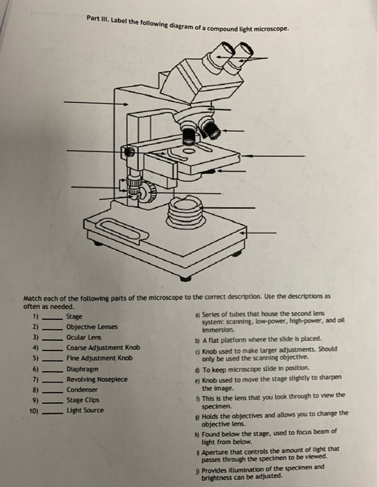
Solved Part III. Label the following diagram of a compound ...

Types of Microscopes: Definition, Working Principle, Diagram ...

Compound Microscope Parts – Labeled Diagram and their ...

Compound microscope labeling Diagram | Quizlet

A labeled diagram of a microscope. MLT 101. :) | Teaching ...

File:Microscope compound diagram.png - Wikimedia Commons
Parts of a Light Microscope Activity | Labeling Task

Dissecting Stereo Microscope Parts and Functions
Parts of a Microscope with Their Functions • Microbe Online
microscope | Types, Parts, History, Diagram, & Facts | Britannica
Label the Light Microscope - Diagram berlabel

Microscope labeling

LABELING THE COMPOUND LIGHT MICROSCOPE 2 Diagram | Quizlet

Compound Microscope Parts, Functions, and Labeled Diagram ...
Post a Comment for "39 light microscope diagram with labels"