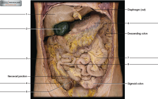
38 label the features of the stomach and nearby regions in this frontal section of a cadaver
Lateral view of the brain: Anatomy and functions | Kenhub These are the superior and inferior frontal sulci. These sulci divide this region into superior, middle, and inferior frontal gyri. The inferior frontal gyrus is further divided into three parts by the stem of the lateral sulcus. These three parts include: The orbital part The triangular part The opercular part The Cerebrum - Lobes - Vasculature - TeachMeAnatomy Central sulcus - groove separating the frontal and parietal lobes. Lateral sulcus - groove separating the frontal and parietal lobes from the temporal lobe. Lunate sulcus - groove located in the occipital cortex. The main gyri are: Precentral gyrus - ridge directly anterior to central sulcus, location of primary motor cortex.
The Abdomen (Human Anatomy) - Picture, Function, Parts ... - WebMD In the front, the abdomen is protected by a thin, tough layer of tissue called fascia. In front of the fascia are the abdominal muscles and skin. In the rear of the abdomen are the back muscles and...

Label the features of the stomach and nearby regions in this frontal section of a cadaver
Serosa four layers of the wall fig 5416 the mucus - Course Hero FIGURE 54.17 Label the features of the stomach and nearby regions in this frontal section of a cadaver (anterior view). A 2 FIGURE 54.18 Label the features associated with the liver and pancreas (liver is removed). A 2 595 mar98655_ch54_581-598.indd 595 11/20/14 10:16 AM 1.4 Anatomical Terminology - Anatomy & Physiology Figure 1.4.1 - Regions of the Human Body: The human body is shown in anatomical position in an (a) anterior view and a (b) posterior view. The regions of the body are labeled in boldface. A body that is lying down is described as either prone or supine. Prone describes a face-down orientation, and supine describes a face up orientation. PDF The Cardiovascular System - Pearson nal anterior view and a frontal section. As the ana-tomical areas of the heart are described in the next section, keep referring to Figure 11.3 to locate each of the heart structures or regions.) Chambers and Associated Great Vessels Learning Objectives Trace the pathway of blood through the heart. Compare the pulmonary and systemic circuits.
Label the features of the stomach and nearby regions in this frontal section of a cadaver. Illustrated Anatomy of the Stomach - ThoughtCo The stomach is an organ of the digestive system. It is an expanded section of the digestive tube between the esophagus and small intestine. Its characteristic shape is well known. The right side of the stomach is called the greater curvature and the left the lesser curvature. Female Body Diagram: Parts of a Vagina, Location, Function Vagina: The vagina is a muscular canal that connects the cervix and the uterus, leading to the outside of the body. Parts of the vagina are rich in collagen and elastin, which give it the ability to expand during sexual stimulation and childbirth. Cervix: The cervix is the lower part of the uterus that separates the lower uterus and the vagina ... Lumen inferior end 544 figure 5411 normalappendix 2 - Course Hero Locate the four layers of the wall (fig. 54.13). The mucus functions as a lubrication and holds the particles of fecal matter together. 5. Prepare a labeled sketch of the wall of the large intestine in Part A of the laboratory assessment. 6. Complete Parts B, C, and D of the laboratory assessment. PDF lab Rep~3-t Organization of the Body - srvhs.org Nume: Date: Section: lab Rep~3-t 2 Organization of the Body Figure 1-3 Figure 1-4 1. - 2. - 3. Multiple Choice 6. Figure 1-5 Multiple Choice (only one response 15 correct In each ~tem) 1. ,4n anatomist cuts a cadaver reserved bodv) with a large raw in a way that divides the cadaver into equal left and right halves.The cut is along a -L planc. a.
Lab 8: Digestive System - Human Anatomy Lab Manual The peritoneum is a large serous membrane which lines the abdominal cavity and coverers most of the digestive organs. some organs are only partially covered by the peritoneum while others are entirely uncovered. These organs are referred to as being retroperitoneal. Body Cavities and Membranes: Labeled Diagram, Definitions Ventral (Anterior) and Dorsal (Posterior) Definitions: Ventral means front or toward the front of the body, and dorsal means back or toward the back of the body. We also learned in the medical prefix lecture that " ventri- " means stomach, abdomen, toward the front, or the anterior aspect of the body. Label the features of the stomach and nearby regions in this frontal ... Mar 22, 2018 · The stomach is divided into four parts: 1-The cardia (8) - this part is connected to the esophagus and its where the epithelium changes from stratified squamous to columnar. In this region is the lower esophageal sphincter (6). 2--The fundus (7)- It's formed by the upper curvature of the stomach. 3- the body (9)- is the main part; and the biggest Abdomen Anatomy, Area & Diagram | Body Maps - Healthline The major muscles of the abdomen include the rectus abdominis in front, the external obliques at the sides, and the latissimus dorsi muscles in the back. The major organs of the abdomen include the...
PDF Body Organization and Terminology ASSIGNMENT Human Cadaver section, click on the appropriate organ system, then click on Show Labels and find: a. External abdominal oblique muscle (Muscular System) b. Sciatic nerve (Nervous System) c. Pancreas and Thyroid gland (Endocrine System) d. Inferior Vena Cava (Cardiovascular System) e. Lymph Vessel (Lymphatic System) f. Appendicular Skeleton (126 bones) | SEER Training Appendicular Skeleton (126 bones) Pectoral girdles. Clavicle (2) Scapula (2) Upper Extremity. Humerus (2) Radius (2) Ulna (2) Carpals (16) Metacarpals (10) Phalanges (28) A and P lab Final part II Flashcards - Quizlet region of stomach near lower esophageal sphincter. Root Canal. contains blood vessels and nerves in a tooth ... Label 54.19 Features of the giestive structures of this abdominopelvic cavity of a cadaver ( anterior view) ... Gallbladder 3. Greater omentum 4. cecum 5.appendix 6. stomach 7. large intestine 8.small intestine. label 54.20 features ... Chart of Major Muscles on the Front of the Body with Labels Sternocleidomastoid. is a paired muscle in the superficial layers of the front part of the neck. It tilts the head to its own side and rotates the head so the head faces the opposite side. It is also an accessory muscle of breathing out and raises the sternum.
Solved Label the anatomical features of the stomach and - Chegg Question: Label the anatomical features of the stomach and nearby regions in the frontal section of a cadaver (anterior view) by clicking and dragging the labels to the correct location tower esophageal sphincter Body of stomach Pancreatic duct Pyloric region Bile duct Fundus of stomach Esophagus Cardia region This problem has been solved!
Post a Comment for "38 label the features of the stomach and nearby regions in this frontal section of a cadaver"